
Page 520 - Adams and Stashak's Lameness in Horses, 7th Edition
P. 520
486 Chapter 4
Treatment
VetBooks.ir Incomplete Avulsion (Coronary Band Not Involved)
Incomplete avulsions of the hoof wall at the heel and
quarter without involvement of the coronary band are
usually best removed. Attempts to salvage the hoof
28
wall are often unrewarding and contribute to continued
lameness and infection beneath the avulsed hoof wall
(Figure 4.61). Removal also prevents the wall from
being snagged and continually traumatized, which is
often very painful. 3
The hoof wall can be removed with the horse stand-
ing for limited involvement or under general anesthesia
for larger lesions. Sharp hoof knives, nippers, and a
handheld electric drill (Dremel tool) to burr the hoof
wall at its attachments may be used for removal. 28,31 The
dorsal attachment of the unaffected hoof wall should be
beveled flush to the wound so there is little tendency for
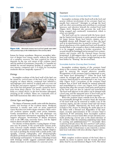
Figure 4.60. Abnormal coronary band and hoof growth associated it to be snagged, resulting in further separation. The
with a previous injury to the medial aspect of the coronary band. wound should be bandaged and protected from contam-
ination and trauma until the exposed tissues become
horses for future soundness. However, secondary infec- keratinized. A full‐support shoe can be used to provide
tion of deeper hoof tissues usually reduces the chances hoof wall stability and to reduce weight‐bearing on the
of a complete recovery. The time required for healing hoof defect by “floating” the involved heel.
depends on the size and extent of the avulsion injury
and the method of treatment. Generally, 3–5 months are Incomplete Avulsion (Coronary Band Involved)
needed for second intention healing of complete avul-
sion injuries, compared with 3–4 weeks for incomplete Incomplete avulsion injuries of the coronary band
avulsions that are surgically repaired. 21,28 alone or the coronary band and hoof wall are best man-
aged by suturing the wound whenever possible.
Etiology Reapposition of the coronary band is important to pre-
vent future hoof deformities. 26,27 Often the hoof wall
Incomplete avulsion of the hoof wall of the heel can cannot be salvaged and will require removal, but the
be caused by vertical tears of the hoof wall, kicking or coronary band should be reconstructed in any way pos-
stepping on sharp objects, continued foot imbalance, sible (Figure 4.62). Lacerations of the coronary band
and improper shoe removal in which nails are torn out without loss of hoof wall should be sutured primarily
of the heel and quarter regions. 28,31 Other avulsion inju- and immobilized in a foot cast (Figure 4.63). Avulsion
ries of the foot and pastern are usually caused by lacera- injuries that affect the coronary band and a small portion
tions from sharp objects. The horse either steps on or of the hoof wall can also be sutured and immobilized.
kicks at a sharp object, or the foot becomes entrapped, The hoof wall adjacent to the defect can be thinned with
resulting in the avulsion. These are commonly seen as a hoof rasp, and the separated piece of hoof wall can be
heel bulb lacerations that often involve the hoof. thinned with a motorized burr to permit suturing. 28,31
When the avulsion injury extends from the solar sur-
Clinical Signs and Diagnosis face proximally through the coronary band, the majority
of the hoof wall can be removed to within 1 cm of the
The degree of lameness usually varies with the duration, coronary region, and the coronary band and soft tissues
extent, and location of the avulsion injury. Moderate sutured if possible. Alternatively, the hoof wall can be
lameness is usually seen with an acute superficial included in the closure by thinning the walls adjacent to
injury that does not involve deeper structures. More exten- the defect with a Dremel tool. Regardless of the tech-
sive avulsion injuries usually cause severe lameness. nique, accurate approximation of the coronary band is
Gentle manipulation of the foot and phalanges can important. If left untreated, these incomplete avulsion
provide important information regarding the status of injuries of the coronary band often remain elevated, even-
support structures. Involvement of deeper structures tually producing a horny spur at the distal extremity of
such as the DIP joint, navicular bursa, and the digital the avulsion, while the remaining underlying tissue heals
tendon sheath should be identified. 3 by scarring and epithelialization. 26,28,31 Invariably these
For chronic avulsion injuries, varying degrees of lame- avulsions protrude above the skin and hoof wall surface,
ness may be present. If the wound heals without prob- making them susceptible to further trauma and painful to
lems, lameness usually subsides with time. However, if palpation. If the avulsed tissue is just removed, a perma-
lameness and purulent exudate persist, further diagnos- nent hoof wall defect will often develop (Figure 4.60).
tics such as probing the wound with a sterile probe, radi-
ography, contrast radiography, and ultrasonography Complete Avulsion
should be performed to determine the cause of the contin-
3
ued drainage and lameness. A chronic nonhealing wound With complete avulsion injuries of the hoof, there is
with drainage usually suggests continued infection of usually no tissue to appose and the wound and hoof
deeper structures or involvement of a synovial cavity. defect heal by second intention (Figure 4.64). The initial