
Page 558 - Adams and Stashak's Lameness in Horses, 7th Edition
P. 558
524 Chapter 4
The cause of simple axial fractures is unknown but of the pastern, and crepitus may be felt but is not a con-
may be associated with repetitive trauma. Comminuted sistent finding. The pastern may also appear to be unsta-
VetBooks.ir a combination of compression and torsion (twisting) just above the coronary band in horses with commi-
ble during manipulation, and swelling may be present
fractures are thought to result from external trauma or
nuted fractures (due to effusion of the DIP joint). With
forces that occur with sudden stops, starts, and short
turns. Most comminuted P2 fractures are thought to biaxial eminence fractures the swelling is less evident
occur as a single‐event injury, but a history of lameness and may not be apparent.
in the affected limb may precede the fracture in some
horses. Horses shod with heel calks are believed to be
more prone to comminuted P2 fractures because the Diagnosis
calks grip the ground, preventing the normal rotation of A definitive diagnosis requires a complete radiographic
the foot and phalanges when the horse rapidly changes examination. At least four views are recommended:
directions. These fractures may also occur in horses dur- dorsopalmar (DP), lateromedial (LM), dorsolateral to
ing light work or unrestrained paddock/pasture exercise palmaromedial oblique (DLPMO), and dorsomedial
due to sudden excessive forces (compression and tor- to palmarolateral oblique (DMPLO). Osteochondral frac-
sion) placed on the limb (“bad step”). Horses turned out tures are usually easily diagnosed with the routine radio-
for exercise after long‐term confinement have also been graphic views. Additional views may be necessary with
reported to be at risk for comminuted P2 fractures. comminuted fractures so that the fracture location and
configuration can be accurately appreciated. Identification
Clinical Signs of whether the fracture lines extend into the DIP joint and
whether there is an intact “strut” of bone that extends
The clinical signs associated with P2 fractures that do between the PIP and DIP is very important information
not disrupt the weight‐bearing capabilities of P2 (osteo- for comminuted fractures. The fracture configuration has
chondral fragments, single eminence, and simple axial considerable bearing on the treatment method selected as
fractures) can be variable. Some horses may have a his- well as the prognosis for future soundness.
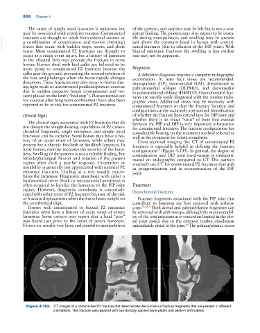
tory of an acute onset of lameness, while others may Cross‐sectional imaging like CT of comminuted P2
present for a chronic forelimb or hindlimb lameness. In fractures is especially helpful in defining the fracture
most horses, exercise increases the severity of the lame- configuration (Figure 4.103). In general, the degree of
57
ness. Swelling of the pastern is not a reliable finding, but comminution and DIP joint involvement is underesti-
fetlock/phalangeal flexion and rotation of the pastern mated on radiographs compared to CT. The authors
region often elicit a painful response. Crepitation or routinely use CT for comminuted P2 fractures that aids
instability is generally not appreciated with uniaxial P2 in prognostication and in reconstruction of the DIP
eminence fractures. Circling at a trot usually exacer- joint.
bates the lameness. Diagnostic anesthesia with either a
basisesamoid nerve block or intrasynovial anesthesia is
often required to localize the lameness to the PIP joint Treatment
region. However, diagnostic anesthesia is contraindi-
cated with other types of P2 fractures because of the risk Osteochondral Fractures
of fracture displacement when the horse bears weight on Fracture fragments associated with the PIP joint that
the anesthetized digit. contribute to lameness are best removed with arthros-
Horses with comminuted or biaxial P2 eminence copy. 52,70,72 Both dorsal and palmar/plantar fragments can
fractures often have a history of acute onset of severe be removed with arthroscopy, although the maneuverabil-
lameness. Some owners may report that a loud “pop” ity of the instrumentation is somewhat limited in the dor-
was heard just prior to the onset of severe lameness. sal joint pouch due to the extensor tendon attachment
Horses are usually very lame and painful to manipulation immediately distal to the joint. The palmar/plantar recess
60
Figure 4.103. CT images of a comminuted P2 fracture that demonstrate the numerous fracture fragments that are present in different
orientations. This fracture was repaired with two dorsally applied bone plates and pastern arthrodesis.