
Page 584 - Adams and Stashak's Lameness in Horses, 7th Edition
P. 584
550 Chapter 4
both sesamoids may be affected. Some lesions appear (inflammation of the synovial membrane), capsulitis
cystic, whereas others appear to erode the axial border (inflammation of the fibrous joint capsule), cartilage
VetBooks.ir Treatment and Prognosis fractures. 9–11,57 Any type of traumatic joint injury can
injury/damage, subchondral bone injury, or intra‐articular
more diffusely.
progress to OA within the fetlock.
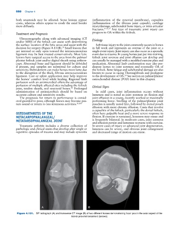
Ultrasonography along with advanced imaging (CT
and/or MRI) of the fetlock can assist with determining Etiology
the surface location of the lytic areas and assist with the Soft tissue injury to the joint commonly occurs in horses
decision for surgery (Figure 4.131B). Small lesions that in full work and represents an overuse of the joint or a
51
are internal or only open toward the intersesamoidean single‐event injury. Joint injury can also occur as a sporadic
ligament may be best treated conservatively. Most lytic event due to trauma. In young horses just put into training,
lesions have surgical access to the area from the palmar/ fetlock joint soreness and joint effusion can develop and
plantar fetlock joint and/or digital sheath using arthros- can usually be managed with a modified exercise plan and
copy. Abnormal bone and ligament should be debrided medication. Abnormal limb conformation may also pre-
if present, and samples are submitted for culture and dispose horses to joint soreness and eventually OA of
sensitivity. Debridement can make horses more lame due the fetlock. Bone fatigue and subchondral damage are also
to the disruption of the thick, fibrous intersesamoidean known to occur in racing Thoroughbreds and predispose
ligament. Cast or splint application may help improve to the development of OA. See section on palmar/plantar
3,4
the horses’ comfort level while healing. Regional limb osteochondral disease (POD) later in this chapter.
perfusion with an antimicrobial offers the advantage of
perfusion of multiple affected sites including the fetlock Clinical Signs
joint, tendon sheath, and sesamoid bones. Prolonged
80
administration of antimicrobials should be based on In mild cases, joint inflammation occurs without
accurate culture and sensitivity results. lameness and is noted as joint soreness on flexion and
The prognosis for return to performance is consid- joint effusion in a young, recently worked or maximally
ered guarded to poor, although horses may become pas- performing horse. Swelling of the palmar/plantar joint
ture sound or return to less strenuous activities. 51,103 pouches is usually noted first, followed by dorsal pouch
effusion with more chronic effusion. Cases that involve
a capsulitis of the fetlock, particularly the dorsal fetlock,
OSTEOARTHRITIS OF THE often have palpable heat and a more severe response to
METACARPOPHALANGEAL/ flexion. If exercise is sustained, lameness may ensue and
METATARSOPHALANGEAL JOINT is frequently bilateral. In moderate cases, joint soreness
and effusion persist and lameness worsens with exercise.
Traumatic arthritis includes a diverse collection of In severe cases of injury or advanced joint degeneration,
pathologic and clinical states that develop after single or lameness can be severe, and obvious joint enlargement
repetitive episodes of trauma and may include synovitis and decreased range of motion can ensue.
A B
Figure 4.131. DP radiograph (A) and transverse CT image (B) of two different horses demonstrating focal lysis in the axial aspect of the
lateral proximal sesamoid (arrows).