
Page 792 - Adams and Stashak's Lameness in Horses, 7th Edition
P. 792
758 Chapter 5
greater trochanter is thought to cause shearing forces
across the physis, resulting in displacement between the
VetBooks.ir Clinical Signs
epiphysis and metaphysis.
There is usually a history of trauma with an immedi-
ate severe lameness. However, some foals are still able to
bear some weight but often stand with a toe‐out, hock‐
in appearance. Swelling and pain over the hip, pelvic
asymmetry, gluteal muscle atrophy, and crepitus with
hip manipulation may be present. However, these signs
were found inconsistently in a report of 25 foals with
capital physeal fractures. 41
Diagnosis
Because of the inconsistency of clinical signs, radiog-
raphy of the hip is necessary for an accurate diagnosis.
Separation of the epiphysis and metaphysis of the femo-
ral neck confirms the diagnosis (Figures 5.143 and
5.145). However, other radiographic abnormalities are
often found, such as comminution of the epiphysis,
greater trochanter or acetabular fracture, or coxofemo-
ral joint luxation/subluxation. Concurrent radio-
15
graphic findings often make potential surgical repair
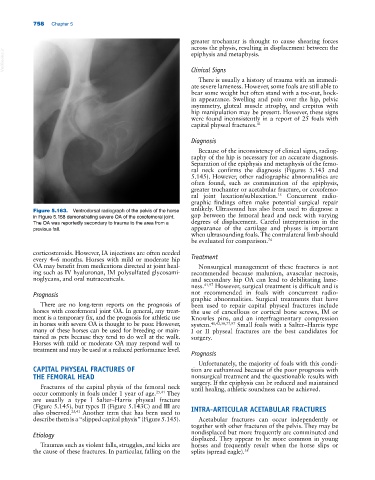
Figure 5.163. Ventrodorsal radiograph of the pelvis of the horse unlikely. Ultrasound has also been used to diagnose a
in Figure 5.158 demonstrating severe OA of the coxofemoral joint. gap between the femoral head and neck with varying
The OA was reportedly secondary to trauma to the area from a degrees of displacement. Careful interpretation in the
previous fall. appearance of the cartilage and physes is important
when ultrasounding foals. The contralateral limb should
be evaluated for comparison. 74
corticosteroids. However, IA injections are often needed
every 4–6 months. Horses with mild or moderate hip Treatment
OA may benefit from medications directed at joint heal- Nonsurgical management of these fractures is not
ing such as IV hyaluronan, IM polysulfated glycosami- recommended because malunion, avascular necrosis,
noglycans, and oral nutraceuticals. and secondary hip OA can lead to debilitating lame-
ness. 41,97 However, surgical treatment is difficult and is
Prognosis not recommended in foals with concurrent radio-
graphic abnormalities. Surgical treatments that have
There are no long‐term reports on the prognosis of been used to repair capital physeal fractures include
horses with coxofemoral joint OA. In general, any treat- the use of cancellous or cortical bone screws, IM or
ment is a temporary fix, and the prognosis for athletic use Knowles pins, and an interfragmentary compression
in horses with severe OA is thought to be poor. However, system. 41,42,50,77,97 Small foals with a Salter–Harris type
many of these horses can be used for breeding or main- I or II physeal fractures are the best candidates for
tained as pets because they tend to do well at the walk. surgery.
Horses with mild or moderate OA may respond well to
treatment and may be used at a reduced performance level.
Prognosis
Unfortunately, the majority of foals with this condi-
CAPITAL PHYSEAL FRACTURES OF tion are euthanized because of the poor prognosis with
THE FEMORAL HEAD nonsurgical treatment and the questionable results with
surgery. If the epiphysis can be reduced and maintained
Fractures of the capital physis of the femoral neck until healing, athletic soundness can be achieved.
occur commonly in foals under 1 year of age. 25,41 They
are usually a type I Salter–Harris physeal fracture
(Figure 5.145), but types II (Figure 5.143C) and III are INTRA‐ARTICULAR ACETABULAR FRACTURES
also observed. 25,41 Another term that has been used to
describe them is a “slipped capital physis” (Figure 5.145). Acetabular fractures can occur independently or
together with other fractures of the pelvis. They may be
Etiology nondisplaced but more frequently are comminuted and
displaced. They appear to be more common in young
Traumas such as violent falls, struggles, and kicks are horses and frequently result when the horse slips or
the cause of these fractures. In particular, falling on the splits (spread eagle). 38