
Page 333 - Feline diagnostic imaging
P. 333
21.2 Contrast adiography 341
(b)
(a)
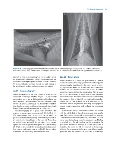
Figure 21.8 Esophagography with iodinated contrast media in a cat with an esophageal mass (arrows). The contrast outlines the
irregular lumen but there is no evidence of leakage of contrast from the esophagus. (a) Lateral projection. (b) Ventrodorsal projection.
stomach as for a pneumogastrogram. The procedure is use- 21.2.10 Barium Enema
ful for evaluation of gastric foreign bodies or neoplasia and
ulceration involving the gastric mucosa. As with a complete The barium enema is a complex procedure that requires
anesthesia and has been largely replaced by endoscopy and
GI series, iodinated contrast should be used instead of
barium if gastric perforation is suspected or imminent. ultrasonography. Additionally, the colon must be thor-
oughly cleansed before the examination. Food should be
withheld for 24 hours and laxatives and enemas should be
21.2.9 Pneumocolography
given the evening before the examination. At 1–2 hours
Pneumocolography is the most common procedure for before the contrast study, a warm water enema should be
evaluation of the large intestine (Figure 21.9). Its primary administered until the expelled fluid is clear. Administering
applications are to aid in differentiation of the large and the enema too close to the study can result in the introduc-
small intestine and evaluation of ileocolic intussusception tion of gas and fluid artifacts. As with other studies, the
or cecal inversion. Although it can be used for identifica- procedure should be preceded by survey radiographs to
tion of colonic masses or strictures, these conditions should ensure proper preparation and evaluate for preexisting
be confirmed with ultrasonography or endoscopy. lesions.
Pneumocolography is a quick, easy procedure that To perform the study, a Foley catheter should be inserted
requires only a syringe or rubber ear bulb filled with air [3]. with the cuff locked in the pelvic canal. A syringe can be
If a portosystemic shunt is suspected, the cat should be used if the patient is too small to accommodate a catheter.
placed in left lateral recumbency to lessen the possibility of Liquid barium suspension (30% w/v) is diluted 1 : 1 with
air embolism in the right side of the heart [3]. The syringe warm water before being placed in an enema bag. The bag
tip is placed in the anus with the front of the barrel pressed is attached to an IV pole to allow the barium to flow by
against the anus to prevent leakage of air. Approximately gravity into the colon at a dosage of 5–15 mL per pound. If
20–30 mL or air is instilled to moderately distend the colon. available, the colon should be watched on fluoroscopy
In a normal study, gas should smoothly fill the ascending, while the barium runs in. Otherwise, a partial dose can be
transverse, and descending portions of the colon. given and then the colon can be evaluated by exposing a