
Page 354 - Feline diagnostic imaging
P. 354
362 22 Gastrointestinal Disease
(a) (c)
(b)
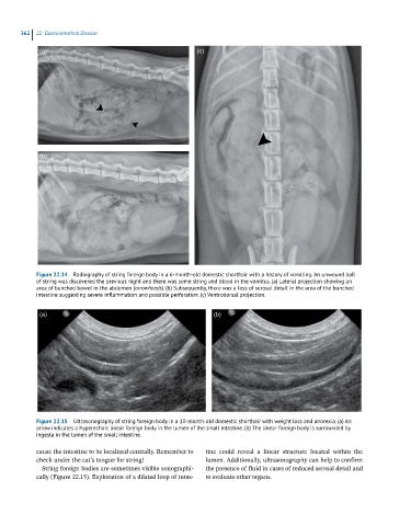
Figure 22.14 Radiography of string foreign body in a 6-month-old domestic shorthair with a history of vomiting. An unwound ball
of string was discovered the previous night and there was some string and blood in the vomitus. (a) Lateral projection showing an
area of bunched bowel in the abdomen (arrowheads). (b) Subsequently, there was a loss of serosal detail in the area of the bunched
intestine suggesting severe inflammation and possible perforation. (c) Ventrodorsal projection.
(a) (b)
Figure 22.15 Ultrasonography of string foreign body in a 10-month-old domestic shorthair with weight loss and anorexia. (a) An
arrow indicates a hyperechoic linear foreign body in the lumen of the small intestine. (b) The linear foreign body is surrounded by
ingesta in the lumen of the small intestine.
cause the intestine to be localized centrally. Remember to tine could reveal a linear structure located within the
check under the cat’s tongue for string! lumen. Additionally, ultrasonography can help to confirm
String foreign bodies are sometimes visible sonographi- the presence of fluid in cases of reduced serosal detail and
cally (Figure 22.15). Exploration of a dilated loop of intes- to evaluate other organs.