
Page 364 - Feline diagnostic imaging
P. 364
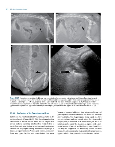
372  22  Gastrointestinal Disease

             (a)                                             (b)


             (c)                                        (d)


            Figure 22.27  Intestinal perforation. An 11-year-old domestic longhair presented with a three-day history of constipation and
            swelling in the perineal region. A diverticulum or perineal hernia (P) was suspected. (a) On the left lateral projection, there was an
            ill-defined area of gas and soft tissue opacity (arrow) associated with the colon in the caudal pelvic cavity. A large amount of
            fecaloid material was present in the rectal area and in the soft tissues caudal to the caudal vertebrae. (b) Right lateral projection.
            (c) Ventrodorsal projection. (d) Ultrasound images showed gas (arrow) that did not appear to be associated with the colon (C).


            22.4.8  Perforation of the Gastrointestinal Tract  because of increased subject contrast between soft tissue and
                                                              gas compared to that seen between soft tissue and normally
            Perforation can result in fluid and/or gas being visible in the   surrounding fat. Gas shapes appear sharp‐edged and have
            peritoneal cavity (Figure 22.27) [51]. On radiographs, free   geometric shapes such as a triangle rather than the rounded
            fluid  causes  a  loss  of  serosal  detail,  which  ranges  from   shapes (ovals, circles) seen with intraluminal gas. Air–fluid
            serosal surfaces appearing indistinct to a complete loss of   interface can be seen if the abdomen is exposed with a hori-
            detail. Free gas is most obvious when it accumulates between   zontal beam and both air and fluid are present. Small bub-
            the liver and diaphragm, causing the liver and diaphragm to   bles  may  be  trapped  in  the  mesentery,  spleen,  or  other
            be seen as separate entities. Where gas is present, serosal sur-  tissues. On ultrasonography, fluid resulting from perforation
            faces  may  appear  brighter  and  more  distinct  than  usual   appears cellular. Free gas results in reverberation artifact.