
Page 376 - Feline diagnostic imaging
P. 376
(b)
(a)
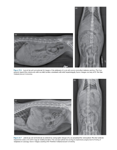
Figure 23.6 Lateral (a) and ventrodorsal (b) images of the abdomen of a cat with poorly controlled diabetes mellitus. The liver
extends beyond the costal arch with rounded borders consistent with mild hepatomegaly. Source: Images courtesy of Dr Merrilee
Holland, Auburn University.
(b)
(a)
Figure 23.7 Lateral (a) and ventrodorsal (b) abdominal radiographic images of a cat presented for constipation. The liver extends
beyond the costal arch with irregular knobbly borders. This cat was euthanized during an exploratory surgery due to finding of
neoplasia on cytology. Source: Images courtesy of Dr Merrilee Holland, Auburn University.