
Page 382 - Feline diagnostic imaging
P. 382
23.6 Abnormal AApmombnp nof tpf pallbp lipo 391
calculated accuracy of ultrasound in the diagnosis of
hepatic lipidosis was only approximately 70% [58]. Diabetes
mellitus can also result in diffuse fatty infiltration of the
liver (Figures 23.20 and 23.21). CCHC in cats can result in
hepatic parenchyma that is normal, hyperechoic, hypo-
echoic, or heterogeneous, and with hepatic size that is
normal or increased (Figure 23.22) [55,59–61]. Biliary
abnormalities. including thickened gallbladder wall and
thickened, dilated, and/or tortuous bile duct. are usually
present as well. Hepatomegaly, hyperechogenicity, and
irregular hepatic margins have been reported with hepatic
amyloidosis in cats [62].
Figure 23.18 Transverse ultrasound image of the liver in a cat
with hepatic lipidosis. The hepatic parenchyma is hyperechoic to
the adjacent ventral falciform fat. The deeper portions of the
liver are attenuated. A lower frequency transducer is needed to
fully image the entire liver.
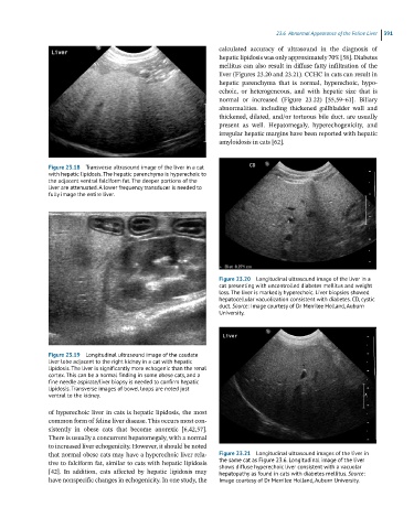
Figure 23.20 Longitudinal ultrasound image of the liver in a
cat presenting with uncontrolled diabetes mellitus and weight
loss. The liver is markedly hyperechoic. Liver biopsies showed
hepatocellular vacuolization consistent with diabetes. CD, cystic
duct. Source: Image courtesy of Dr Merrilee Holland, Auburn
University.
Figure 23.19 Longitudinal ultrasound image of the caudate
liver lobe adjacent to the right kidney in a cat with hepatic
lipidosis. The liver is significantly more echogenic than the renal
cortex. This can be a normal finding in some obese cats, and a
fine needle aspirate/liver biopsy is needed to confirm hepatic
lipidosis. Transverse images of bowel loops are noted just
ventral to the kidney.
of hyperechoic liver in cats is hepatic lipidosis, the most
common form of feline liver disease. This occurs most con-
sistently in obese cats that become anorexic [6,42,57].
There is usually a concurrent hepatomegaly, with a normal
to increased liver echogenicity. However, it should be noted
that normal obese cats may have a hyperechoic liver rela- Figure 23.21 Longitudinal ultrasound images of the liver in
tive to falciform fat, similar to cats with hepatic lipidosis the same cat as Figure 23.6. Longitudinal image of the liver
shows diffuse hyperechoic liver consistent with a vacuolar
[42]. In addition, cats affected by hepatic lipidosis may hepatopathy as found in cats with diabetes mellitus. Source:
have nonspecific changes in echogenicity. In one study, the Image courtesy of Dr Merrilee Holland, Auburn University.