
Page 405 - Feline diagnostic imaging
P. 405
414 24 Pancreas
(a) (b)
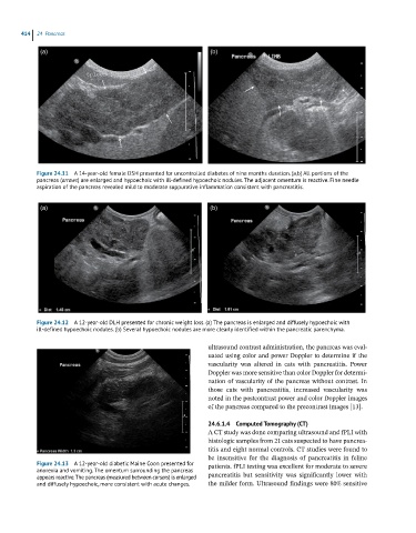
Figure 24.11 A 14-year-old female DSH presented for uncontrolled diabetes of nine months duration. (a,b) All portions of the
pancreas (arrows) are enlarged and hypoechoic with ill-defined hypoechoic nodules. The adjacent omentum is reactive. Fine needle
aspiration of the pancreas revealed mild to moderate suppurative inflammation consistent with pancreatitis.
(a) (b)
Figure 24.12 A 12-year-old DLH presented for chronic weight loss. (a) The pancreas is enlarged and diffusely hypoechoic with
ill-defined hypoechoic nodules. (b) Several hypoechoic nodules are more clearly identified within the pancreatic parenchyma.
ultrasound contrast administration, the pancreas was eval-
uated using color and power Doppler to determine if the
vascularity was altered in cats with pancreatitis. Power
Doppler was more sensitive than color Doppler for determi-
nation of vascularity of the pancreas without contrast. In
those cats with pancreatitis, increased vascularity was
noted in the postcontrast power and color Doppler images
of the pancreas compared to the precontrast images [13].
24.6.1.4 Computed Tomography (CT)
A CT study was done comparing ultrasound and fPLI with
histologic samples from 21 cats suspected to have pancrea-
titis and eight normal controls. CT studies were found to
be insensitive for the diagnosis of pancreatitis in feline
Figure 24.13 A 12-year-old diabetic Maine Coon presented for patients. fPLI testing was excellent for moderate to severe
anorexia and vomiting. The omentum surrounding the pancreas
appears reactive. The pancreas (measured between cursors) is enlarged pancreatitis but sensitivity was significantly lower with
and diffusely hypoechoic, more consistent with acute changes. the milder form. Ultrasound findings were 80% sensitive