
Page 426 - Feline diagnostic imaging
P. 426
436 25 Adrenal Gland
(a) (b)
Figure 25.17 A 7-year-old female DSH presented for icterus following surgical repair of a cruciate rupture a week earlier. She was
diagnosed with acute renal and liver failure following presentation to our clinic. On abdominal ultrasound, the width of the right
adrenal gland is enlarged (a). The length of both adrenal glands appeared increased which may be due to current illness (b).
(a) (b)
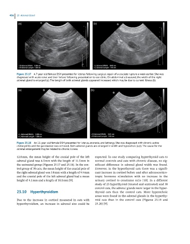
Figure 25.18 An 11-year-old female DSH presented for icterus, anorexia, and lethargy. She was diagnosed with chronic active
cholecystitis and the gallbladder was removed. Both adrenal glands are enlarged in width and hypoechoic (a,b). The cause for the
adrenal enlargement may be related to chronic illness.
12.0 mm, the mean height of the cranial pole of the left expected. In one study comparing hyperthyroid cats to
adrenal gland was 4.5 mm with the length of 11.5 mm in normal controls and cats with chronic disease, no sig -
the untreated group (Figures 25.17 and 25.18). In the con- nificant difference in adrenal gland width was found.
trol group of 30 cats, the mean height of the cranial pole of However, in the hyperthyroid cats there was a signifi-
the right adrenal gland was 3.8 mm with a length of 9.9 mm cant increase in cortisol before and after adrenocortico -
and the cranial pole of the left adrenal gland had a mean tropic hormone stimulation with no increase in the
height of 4.1 mm and a length of 10.1 mm [9]. urinary cortisol to creatinine ratio [10]. In a different
study of 23 hyperthyroid (treated and untreated) and 30
control cats, the adrenal glands were larger in the hyper -
25.10 Hyperthyroidism thyroid cats than the control cats. More hyperechoic
areas were found in the adrenal glands in the hyperthy -
Due to the increase in cortisol measured in cats with roid cats than in the control cats (Figures 25.19 and
hyperthyroidism, an increase in adrenal size could be 25.20) [9].