
Page 447 - Feline diagnostic imaging
P. 447
27.1 he eline idneys 459
(a) (b)
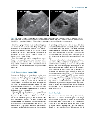
Figure 27.5 Ultrasonography of pyelonephritis in a 16-year-old domestic longhair. (a) Midsagittal image of the left kidney showing
normal length (4.41 cm) but reduced corticomedullary definition and mild pelvic dilation at 0.66 cm. (b) Transverse ultrasound image
of the left kidney showing mild dilation of the renal pelvis and the proximal ureter (0.2 cm between the calipers).
On ultrasonography (Figure 27.6), the distended pelvis of cysts frequently increased between scans [12]. Cats
and diverticula are anechoic with sharp margins and younger than 10 months that are initially negative should
hydroureter is usually present. In severe cases, the con- be reexamined when they mature. Additionally, those that
tents may be anechoic but are usually cellular, causing are positive should be removed from the breeding pool.
the kidney to resemble a large abscess. The hyperechoic Serial ultrasonography can be beneficial in determining
remnants (Figure 27.6c) of the diverticula help to iden- prognosis because the condition has a progressive course
tify the structure as the kidney. [12]. Renal failure is related to the number and size of cysts
Since hydronephrosis implies obstruction, a search [10].
should be conducted to determine the cause, which On survey radiography, the affected kidney may be nor-
includes ureteral calculus, ureteral stricture, mucus mal or large and distorted (Figure 27.7). Cysts can be suf-
plugs, external compression of the ureter, ectopic ureter, ficiently numerous or large to cause the kidney to be
and neoplasia at the bladder trigone or rarely in the ureter mass‐like. On excretory urography, normal parts of the
itself [7, 8]. renal parenchyma opacify but the cysts will be less radio-
paque, appearing as filling defects. On ultrasonography,
the classic cyst is anechoic with well‐defined walls and
27.1.5 Polycystic Kidney Disease (PKD)
deep acoustic enhancement (Figure 27.8). There may be a
Although the incidence of insignificant cortical cysts few or many small cysts (Figure 27.9) but cysts can also
increases with age in dogs and humans, insignificant cysts become very large (Figures 27.7 and 27.10), causing defor-
are uncommon in cats [9]. The incidence of PKD was mation of the renal margins [10]. On occasion, the kidney
investigated in 250 long‐haired and 14 short‐haired may become so distorted that neoplasia is suspected and
(“exotic”) Persian cats showing that 45% of the long‐haired aspiration or biopsy of remaining parenchyma may be nec-
and 50% of the short‐haired cats were affected [10]. Four essary to exclude this possibility. Five percent of Persian
Ragdolls and three British shorthair cats were negative for cats with PKD may have involvement of the liver and pan-
PKD. These findings were consistent with an autosomal creas (Figure 27.11).
dominant hereditary transmission.
Although PKD may be diagnosed in younger kittens, 27.1.6 Neoplasia
another study found that the sensitivity of sonographic
diagnosis was 75% for 16‐week‐old kittens compared to Because renal masses are in the retroperitoneal space,
91% for cats older than 3 years [11]. When 183 cats older they remain in a dorsal location (Figure 27.12). Large
than 10 months (57 Persians, 13 exotic shorthairs, six renal masses may appear to be located in midabdomen
British shorthairs, two Selkirk Rex cats) were scanned with because they grow ventrally to fill the dorsoventral
ultrasonography, 31% were positive for PKD. When 78 cats dimension. Close observation reveals that there is only
were rescanned 11–13 months later, there was agreement fat located dorsal to the mass and other than the mass,
between the results for both scans but the size or number there is no other candidate for the kidney on the side of