
Page 481 - Feline diagnostic imaging
P. 481
28.6 Diseisi of tsf seeas sep odu Dis Si se 493
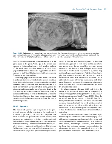
Figure 28.11 Radiography of dystocia in a 1-year old cat. A single live kitten was delivered the night before and no contractions
were seen during the past 2–3 hours. On radiography, there were three fetuses with no evidence of obstruction or fetal death. One
fetal heart beat was detected on ultrasound but fetal positioning prevented assessment of the other two.
dence of healed fractures that compromise the size of the causes a focal or multifocal enlargement rather than
pelvic canal in the queen. Visible gas in the uterus, fetal uniform enlargement of both horns so that the uterus
thoracic or abdominal cavities, or fetal vessels or collapse may appear mass‐like or resemble a pregnant uterus.
of the skull bones are clear evidence of fetal death Remember that the uterus must be larger than the small
(Figure 28.12). Loss of flexion also suggests fetal death but intestine to be identified; early cases of pyometra may
this sign by itself should be interpreted with care because a not be radiographically apparent. Additionally, radiogra
fetus may be merely stretching. phy just shows enlargement of the uterus. Physical
Ultrasonography can be used to check the fetal heart rate examination, blood tests, and ultrasound can be used to
to make sure that it is not too fast or too slow. A heart rate differentiate causes of uterine enlargement and deter
below 180 bpm indicates an emergency situation. A lack of mine if pyometra is present. Pregnancy and other causes
movement and no detectable heart beat are proof that fetal of fluid in the uterus such as mucometra and hydrome
death has occurred. Excessive fluid in uterus, gas in the tra must be considered.
uterus or fetal tissues, and a loss of expected detail in the On ultrasonography (Figures 28.13 and 28.14), the
fetal anatomy are further evidence of fetal death. Rarely, a lumen of the uterus will be anechoic or echogenic [10]
mummified fetus may be seen in the abdomen. If the fetus with a swirling pattern although the type of fluid cannot
has been dead for some time, it may become a mineralized be determined sonographically (Figure 28.15). Often, the
mass in which the bones are compressed and the fetus is uterus will have distinct, multifocal fluid‐filled swellings
barely recognizable. throughout both uterine horns. The uterus should not be
aspirated transabdominally to avoid spilling purulent
material into the peritoneal cavity. When infection occurs
28.6.2 Pyometra
in the uterine stump remaining after ovariohysterectomy,
The classic radiographic sign of pyometra is the pres a fluid‐filled structure will be seen between the bladder
ence of tortuous tubular opacities in the caudoventral and colon.
abdomen (Figure 28.13). On the lateral projection, the If uterine rupture occurs, the abdomen will exhibit a par
small intestines are pushed dorsally and cranially and tial or complete loss of serosal detail on radiography. Other
the colon and bladder may be further apart than normal. differentials include rupture of another hollow organ such
In less obvious cases, adjacent organs can be compressed as the urinary bladder, ascites associated with cardiac or
and separated using a wooden spoon or commercial pad liver disease, hemorrhage, peritonitis, and neoplasia
dle to improve visualization of the uterus. The tortuous involving the peritoneal surface (carcinomatosis). On
fluid‐opaque horns are more difficult to appreciate on ultrasonography, echogenic material will be seen in the
the ventrodorsal projection. Pyometra in the queen often peritoneal space similar to the fluid seen in the uterus.