
Page 508 - Feline diagnostic imaging
P. 508
520 29 Hemolymphatic System
minerals, iron is hyperechoic. Iron deposits have been
described in the feline spleen [28], suggesting that hemosi-
derosis in the feline spleen would cause speckles similar to
those described in dogs. These are most often seen in
Cushing disease, diabetes mellitus, chronic steroid use,
and immune‐mediated hemolytic anemia.
Other differentials for focal, hyperechoic nodules include
nodular hyperplasia, extramedullary hematopoiesis, neo-
plasia, granulomas, and infarction. The most common tar-
get of the visceral form of mast cell tumor is the spleen and
this tumor comprises around 15–26% of splenic neoplasms
[10,21]. Mast cell tumors (Figure 29.30) can be either
Figure 29.24 Ultrasound scan of a 12-year-old domestic shorthair
with a one-month history of weight loss diagnosed with disseminated hypo‐ or hyperechoic but hyperechoic nodules are more
histoplasmosis. The spleen was mildly enlarged and diffusely mottled likely to be mast cell tumors than lymphoma. Peritoneal
with hypoechoic areas (arrows) scattered throughout.
(a) (b)
Figure 29.25 Ultrasonography of extramedullary hematopoiesis. (a) Image of a 12-year-old Himalayan cat with biliary cystadenoma. The spleen
was enlarged on ultrasonography. Cytology of splenic aspirates obtained during ultrasound showed extramedullary hematopoiesis. (b) Ultrasound
image of a 14-year-old domestic longhair with chronic renal disease and immune-mediated hemorrhagic anemia. The spleen was enlarged
with a hyperechoic nodule. An arrow indicates the tip of a needle as it approaches the nodule. Cytology showed extramedullary hematopoiesis.
effusion and lymphadenopathy are more common in
lymphoma than mast cell tumor [10]. Hemangiosarcomas
may also be hyperechoic.
As noted previously, a common presentation of both
lymphoma and mast cell tumor in the spleen is the pres-
ence of hypoechoic nodules which give the spleen a
“moth‐eaten” or mottled appearance. Hypoechoic nodules
in the spleen, liver, and kidney and lymphadenopathy are
frequent sonographic findings in malignant histiocytosis in
dogs [29]. There are sparse reports of the ultrasound
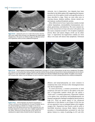
Figure 29.26 Ultrasonography of lymphoid hyperplasia. A appearance of this disease in cats (Figure 29.29c) but one
10-year-old Ocicat was presented for removal of bladder stones. cat was reported to have an enlarged spleen with irregular
On ultrasonography, in addition to changes in the urinary margins [30]. Hypoechoic focal lesions could be solid or
bladder, there was diffuse thickening of the intestinal wall, one fluid‐filled structures (viscous fluid). Splenic infarction
enlarged mesenteric lymph node, and a mottled appearance to
the spleen. Ultrasound-guided aspirates of the spleen and secondary to thromboembolism accounted for only four
subsequent surgical biopsy showed lymphoid hyperplasia. splenic lesions in a study of pathology in 455 cats [19].