
Page 514 - Feline diagnostic imaging
P. 514
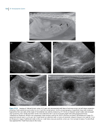
526  29  Hemolymphatic System

                                   (a)


             (b)                                              (c)


             (d)                                        (e)


            Figure 29.34  Imaging of infected lymph nodes. A 1.5-year-old cat presented with fever of unknown origin. (a) Left lateral projection
            showing a fluid opacity (arrows) ventral to L6, L7, and the cranial sacrum. (b) On ultrasonography, a hypoechoic mass with scattered
            hyperechoic foci was seen at the origin of the external iliac arteries, measuring 2.9 × 4.8 cm. (c) Adjacent lymph nodes were enlarged
            and hypoechoic. Fine needle aspiration of the mass indicated that it was an enlarged lymph node with pyogranulomatous
            inflammation. Peritoneal effusion was polymerase chain reaction positive for feline infectious peritonitis. (d) Ultrasound image of a
            mesenteric lymph node in a 16-year-old mixed-breed cat admitted with a history of anorexia. Calipers measure one node at 2.1 cm.
            (e) Ultrasound image of additional nodes in the cat in (d). Some areas are anechoic; others are more cellular in nature. Purulent fluid
            was aspirated from fluid-filled areas in the nodes.