
Page 1279 - Cote clinical veterinary advisor dogs and cats 4th
P. 1279
644 Melanoma
Melanoma Bonus Material Client Education
Online
Sheet
VetBooks.ir Etiology and Pathophysiology ○ Tumors with a mitotic index of < 3/10
BASIC INFORMATION
• Underlying genetic mutations and ultraviolet high-power fields (HPF) are typically
Definition light exposure are known etiologic agents in benign; those ≥ 3/10 HPF are generally
A common neoplasm that develops from humans. malignant.
melanocytes in dogs (rare in cats). Classification • Melanomas do not have to arise in pigmented
of melanomas by anatomic location generally skin. TREATMENT
has more clinical utility than by histologic
subtype. DIAGNOSIS Treatment Overview
• Most oral melanomas are malignant, locally The treatment plan depends on the progno-
invasive, and highly metastatic (>60%) Diagnostic Overview sis, and staging to identify local and meta-
(p. 714). Biopsy of an identified mass is the test of static disease is essential. Goals of treatment
• Subungual (nail bed) melanomas are locally choice to establish a diagnosis of melanoma. A are long-term disease control in patients
invasive with a lower rate of metastasis diagnostic and treatment approach is presented amenable to definitive therapy, and pallia-
(30%-60%). on p. 1437. tion of clinical signs in patients not treated
• Cutaneous melanomas in dogs are generally definitively or those with metastatic disease
(but not always) benign. Differential Diagnosis (p. 1437).
• Ocular melanoma (p. 559) • Oral: squamous cell carcinoma, fibrosarcoma,
acanthomatous ameloblastoma Acute and Chronic Treatment
Synonyms • Cutaneous: any mass lesion of skin (neo- Oral melanoma:
Malignant melanoma, melanocytic tumor plastic or non-neoplastic) • Radical excision of mass if
(benign and malignant), melanocytoma • Subungual: squamous cell carcinoma, nail ○ Wide surgical margins (>2 cm, including
(benign) bed infection underlying bone) can be obtained, and
○ Patient has no regional lymph node or
Epidemiology Initial Database distant metastasis
SPECIES, AGE, SEX CBC, serum biochemistry profile, urinalysis, • Removal of macroscopic (measurable)
Melanoma generally occurs in older patients lymph node aspiration, thoracic radiographs, tumor plus definitive radiation therapy to
(9-12 years). and for patients with hindlimb or caudally primary tumor site and regional lymph
located masses, abdominal ultrasound nodes if
GENETICS, BREED PREDISPOSITION ○ Radical excision is not possible or regional
• Predisposed dog breeds include chow Advanced or Confirmatory Testing lymph node metastasis is identified, and
chow, Doberman pinscher, golden • Biopsy and histopathologic exam of tissue ○ Patient has no distant metastatic disease
retriever, Gordon setter, Irish setter, giant • Immunohistochemical staining with S-100 • Definitive radiation therapy alone to primary
schnauzer, miniature schnauzer, Scottish or Melan-A may confirm the diagnosis of tumor and regional lymph nodes if
terrier (suggests an underlying genetic melanoma in undifferentiated and amelanotic ○ Removal of macroscopic tumor burden
mechanism) tumors. is not possible, and
• Black dogs may be predisposed, but any color • Mitotic index may help distinguish ○ Patient has no distant metastatic disease
dog may be affected. benign from malignant canine cutaneous • Palliative radiation therapy to tumor could
melanomas. be considered if
Clinical Presentation ○ Determination of mitotic index requires ○ The patient has distant metastatic disease,
HISTORY, CHIEF COMPLAINT histopathologic evaluation of tissue and and/or
Oral melanoma: cannot be reliably assessed on fine-needle ○ Financial or other restrictions preclude
• Detection of an oral mass by a veterinarian aspiration/cytology. definitive therapy
during routine exam or dental prophylaxis
(common)
• Identification of an oral mass by the owner
• Recent-onset halitosis, ptyalism
Cutaneous or subungual melanoma:
• Identification of a mass by the owner or
veterinarian
PHYSICAL EXAM FINDINGS
• Exam typically reveals a mass lesion.
• Oral exam should be thorough; some tumors
are located at the base of the tongue or in
the tonsils.
• Cutaneous and subungual tumors may
become ulcerated.
• Melanomas may be pigmented (≈ 3 ) or
2
amelanotic (≈ 3 ).
1
• Thorough exam of draining lymph nodes is
always indicated; cytologic evaluation should
be performed even if the nodes are of normal
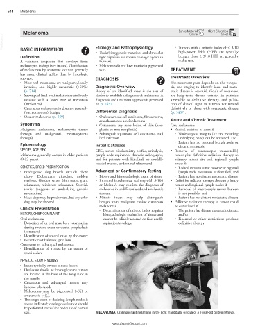
size. MELANOMA Oral malignant melanoma in the right mandibular gingiva of a 7-year-old golden retriever.
www.ExpertConsult.com