
Page 1479 - Cote clinical veterinary advisor dogs and cats 4th
P. 1479
Papillomas, Oral and Cutaneous 753
VetBooks.ir Diseases and Disorders
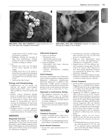
PAPILLOMAS, ORAL AND CUTANEOUS Cutaneous papillomatosis in an PAPILLOMAS, ORAL AND CUTANEOUS Pigmented viral plaques in an
adult cocker spaniel dog. (Copyright Dr. Karen Moriello.) adult pug dog. (Copyright Dr. Karen Moriello.)
medial surfaces of limbs, usually in pugs Differential Diagnosis • Anecdotal reports exist that crushing lesions
and miniature schnauzers • Canine oral papillomas spurs resolution, but this has never been
○ Papillomas affecting the footpads of ○ Fibromatous epulis proved in controlled studies.
dogs. Firm, hyperkeratotic, hornlike ○ Transmissible venereal tumor • Single-case series demonstrated varied
growths that occur on multiple footpads, ○ SCC (especially if ulcerated) response of canine oral papillomatosis to
sometimes resulting in lameness • Cutaneous papilloma azithromycin 5-10 mg/kg PO q 12-48h.
• Cats ○ Pedunculated with fronds: sebaceous • Surgical removal: CO 2 laser ablation, exci-
○ Multiple oral, raised, oval, flat-topped, adenoma/hyperplasia sion, cryosurgery, or electrosurgery. This often
4-8 mm masses, especially on ventral ○ Pigmented: melanoma needs to be repeated. Most important in
tongue ○ Inverted: intracutaneous cornifying cases when normal physiologic functions
○ Variably sized 3-mm to 3-cm masses on epitheliomas are compromised by disease (e.g., airway
haired skin and progress from pigmented obstruction, dysphagia)
macules to hyperkeratotic plaques, which Initial Database • Discontinue use of systemic glucocorticoids,
can transform into the ulcerated lesions Skin biopsy and histologic exam: Classic find- oclacitinib or cyclosporine, particularly if oral
of SCC. ings for exophytic papillomas include epidermal or cutaneous disease reoccurs or persists.
○ Small, pedunculated masses hyperplasia, orthokeratotic hyperkeratosis,
ballooning degeneration of epidermal cells, Chronic Treatment
Etiology and Pathophysiology koilocytosis, variably present intranuclear • Interferon (IFN) at immunostimulatory low
• Papillomaviruses are host specific and inclusion bodies and prominent keratohyaline dosages (0.1 mL of 30,000 IU/mL solution,
relatively site specific, non-enveloped, granules in the stratum spinosum. Exam may = 3000 IU/dose) PO q 24h or high dosages
2
double-stranded DNA viruses that can reveal SCC in situ or invasive SCC (p. 939). (1-1.5 million IU/m SQ q 48-72h [p. 609])
induce proliferative cutaneous and mucosal for 4-8 weeks pending response
tumors. Advanced or Confirmatory Testing • Imiquimod 5% (Aldara) is applied topi-
• Infection requires inoculation through • Most diagnostic laboratories can make a cally to affected areas q 24-72h. Human-
breaks in the epidermal or mucosal barrier definitive diagnosis based on histologic approved immunomodulator that activates
by means of direct contact with an animal findings. PCR, immunohistochemistry, and toll-like receptors upregulates interleukin-6
or transmission by fomite. Immunosup- in situ hybridization can often help support (IL-6), IL-12, tumor necrosis factor-alpha
pression or immunodeficiency may be or confirm a diagnosis. (TNF-alpha), IFN-alpha, and IFN-gamma;
necessary for development of some forms • Electron microscopy is the gold standard for activates Langerhans cells; and provides
(e.g., feline and canine plaques, disseminated diagnosis but is primarily used for research antiproliferative and antiviral effects. Local
papillomatosis). purposes. irritation is anticipated as part of the upregu-
• Incubation period is 1-8 weeks; regression lated immune response against the virus-
typically occurs in 1-5 months, and lesions TREATMENT infected keratinocytes and can sometimes
occasionally persist for 24 months or more. be difficult to differentiate from resolving
Treatment Overview lesions.
DIAGNOSIS • Canine oral papillomatosis, unless severe, • Autogenous vaccines are not recom-
regresses spontaneously and requires no mended because they have been associated
Diagnostic Overview treatment. with injection-site neoplasms several years
For young dogs, diagnosis of canine oral • Consideration of an underlying immunosup- later.
papillomatosis is often visual. All other clinical pressive disease is warranted for adult animals. ○ Modified papillomavirus vaccines contain-
presentations require definitive diagnosis by skin ing codon-optimized E1, E2, E4, and
biopsy and histologic exam of tissue; polymerase Acute General Treatment E7 are protective against experimental
chain reaction (PCR) testing may be required • Lesions often regress spontaneously in 2-3 and natural infection without inducing
for confirmation of diagnosis. months. injection-site neoplasia.
www.ExpertConsult.com