
Page 1585 - Cote clinical veterinary advisor dogs and cats 4th
P. 1585
796 Pneumonia, Bacterial
○ Alveolar pattern, often most severe in
dependent regions of lung lobes, is
VetBooks.ir ■ Right middle lung lobe is commonly
expected in most cases.
affected and may be obscured by the
heart on right lateral projections.
Distribution may be variable in patients
■
that are non-ambulatory or aspirate
after surgery.
○ Interstitial patterns, with or without
alveolar patterns, are possible.
○ In some cases, lobar consolidation is the
prominent radiographic abnormality.
○ Other abnormalities that reflect underlying
primary disease (e.g., megaesophagus,
bronchiectasis, pulmonary mass)
A B
Advanced or Confirmatory Testing
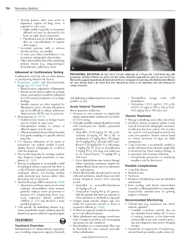
Confirmatory tests help rule out other diseases PNEUMONIA, BACTERIAL A, Left lateral thoracic radiograph of a 14-year-old, mixed-breed dog with
pneumonia. Alveolar infiltrates are visible over the cardiac silhouette (especially the apex) but are not striking.
or predisposing diseases/risk factors: Microcardia suggests hypovolemia. B, Dorsoventral thoracic radiograph of same dog. Left-sided alveolar infiltrates
• Respiratory washes and bronchoalveolar are more obvious than in A, where they were obscured by being on the dependent side and overlying the
lavage (pp. 1073 and 1074) cardiac silhouette.
○ Primarily septic, suppurative inflammation
○ Bacteria are not always evident in cytologic
exams, and samples should be submitted
for C&S testing irrespective of cytologic and addressing predisposing factors to the extent ○ Theophylline: dosage varies with
findings. possible are key. formulation.
○ Specific requests are often required for ○ Terbutaline 1.25-5 mg/DOG PO q 8h;
Mycoplasma culture. Because Mycoplasma Acute General Treatment 0.625-1.25 mg/CAT PO q 12h or 0.01-
spp can be difficult to culture, polymerase Broad-spectrum antibiotics: 0.02 mg/kg SQ or IM once (cat)
chain reaction (PCR) may be preferred. • Frequent in vitro resistance to empirically
• Bronchoscopy (p. 1074): chosen antimicrobials underscores the benefit Chronic Treatment
○ Endobronchial masses or foreign bodies of C&S testing. • Manage underlying causes when identified.
may be evident in some cases. • Clinically unstable animals should be treated • Antibiotic therapy continued at least 1 week
○ Mucopurulent exudate in airways of with broad-spectrum (ideally, parenteral) beyond clinical and radiographic resolution
affected regions may be seen. antibiotics. of infection has been typical, but recently,
○ Dilated/sacculated airways (bronchiectasis) ○ Ampicillin 22-30 mg/kg IV, SQ q 8h, the need for such prolonged treatment has
○ Can guide sampling to a specific affected cefazolin 22 mg/kg IV, IM q 6h, or come into question. Shorter courses based
lung lobe clindamycin 10 mg/kg SQ q 12h (dogs), on clinical resolution of signs are likely
• CT: superior delineation of the extent of 10-15 mg/kg SQ q 12h (cats) AND enro- appropriate.
pneumonia but seldom needed if good- floxacin 5-20 mg/kg IM, IV q 24h (dogs), • Lung lobectomy is occasionally needed to
quality thoracic radiographs are consistent 5 mg/kg IM, IV (cats) or pradofloxacin resolve infection when extensive single-lobe
with the diagnosis 5 mg/kg PO q 24h (dog; extra-label use involvement has failed medical therapy or
• Fine-needle aspiration for cytologic analysis: in the United States), 7.5 mg/kg PO q is associated with recurrent infection.
may diagnose fungal pneumonia or neo- 24h (cats) ○ Foreign-body pneumonia or underlying
plasms (p. 1113) ○ Severe clinical disease may warrant therapy neoplasia may be discovered.
• Barium esophagram is occasionally useful before obtaining respiratory samples for
if esophageal disease suspected, but there is culture; blood culture can be an alternative Possible Complications
a risk of barium aspiration in patients with for such patients. • Lung lobe abscess
esophageal disease. Free-feeding swallow • Mildly affected stable animals may be treated • Bronchiectasis
study protocols may increase utility while with oral antibiotics, ideally based on C&S • Pyothorax
decreasing risk of aspiration. testing. Empirical choices while awaiting • Recurrence if underlying cause not identified
• Arterial blood gas (ABG) analysis (p. 1058) results: and treated
○ Hypoxemia and hypocapnia are the most ○ Ampicillin or amoxicillin/clavulanate • Joint cartilage and dental abnormalities
common abnormalities; when present, 22 mg/kg q 8h, or secondary to fluoroquinolone or tetracycline
generally indicate severe pneumonia or ○ Doxycycline 5 mg/kg PO q 12h, particu- (respectively) administration to growing
presence of a complicating factor (e.g., larly for animals that show no response to puppies or kittens
acute respiratory distress syndrome other antibiotics or Bordetella is suspected.
[ARDS] [p. 27]) and therefore a more • Oxygen (nasal cannula, oxygen cage, face Recommended Monitoring
guarded prognosis. mask) for hypoxemic animals or those in • Clinical signs (e.g., respiratory rate, effort;
• Tests specific for underlying diseases (e.g., respiratory distress (p. 1146) attitude, appetite)
serum acetylcholine receptor antibody titers for • IV fluids are typically needed for moderately ○ Carefully re-evaluate animals that are
myasthenia gravis–induced megaesophagus) or severely affected animals. not clinically better within 48-72 hours
• Saline nebulization and coupage, several times of starting treatment or that deteriorate
TREATMENT daily. Change in position and encouraging substantially at any time; consider pursu-
movement are also beneficial. ing more diagnostic tests and/or changing
Treatment Overview • Bronchodilator use is controversial. They may therapy.
Administration of antimicrobials; supportive be beneficial for some animals, especially • Assessment of oxygenation, if needed (e.g.,
care, including oxygenation support if needed; before nebulization. arterial blood gas analysis, pulse oximetry)
www.ExpertConsult.com