
Page 1708 - Cote clinical veterinary advisor dogs and cats 4th
P. 1708
860 Pythiosis and Lagenidiosis
Pythiosis and Lagenidiosis Bonus Material Client Education
Online
Sheet
VetBooks.ir
PHYSICAL EXAM FINDINGS
BASIC INFORMATION
inguinal lymph nodes, lung, pulmonary
Dog: including great vessels, sublumbar and
Definition • GI infection: severe weight loss in an hilus, and cranial mediastinum.
Oomycotic infections of the gastrointestinal otherwise active, alert dog; abdominal mass
(GI) tract (Pythium) or skin (Pythium, Lagen- often palpable. Colonic mucosa may feel DIAGNOSIS
idium, and Paralagenidium) of dogs and cats thickened/cobblestone on rectal exam.
caused by Pythium insidiosum, Lagenidium spp, • Cutaneous infection: nonhealing wounds Diagnostic Overview
and Paralagenidium karlingii, respectively and invasive masses that contain ulcerated The diagnosis of pythiosis is suspected in one of
nodules and draining tracts, most often two contexts: chronic weight loss with vomiting
Synonyms involving extremities, tailhead, ventral neck, and/or diarrhea (marked intestinal thickening/
Oomycosis, phycomycosis, swamp cancer or perineum mass raises the differential diagnosis of neoplasia)
Cat: or chronic, invasive, often ulcerated subcutaneous
Epidemiology • Nasopharyngeal infection: nasal discharge masses. Confirmation is by serology or histologic
SPECIES, AGE, SEX and upper respiratory signs; ulcerated caudal demonstration of the organism on biopsy. If
Dogs (young to middle-aged) affected more oral masses cutaneous oomycotic infection is noted, systemic
often than cats (any age but often < 1 year) • Cutaneous infection: invasive subcutaneous disease should be suspected in Lagenidium or
masses (especially in inguinal, tailhead, and Paralagenidium infections.
GENETICS, BREED PREDISPOSITION periorbital regions) or ulcerated plaquelike
Dogs: any breed, especially large, outdoor lesions on extremities Differential Diagnosis
working breeds • GI infection: occasionally seen Gastrointestinal presentations:
• GI neoplasia
RISK FACTORS Etiology and Pathophysiology • Other causes of esophageal disease
Recurrent exposure to warm freshwater lakes, • Infection follows exposure of the immuno- • Pyloric outflow obstruction due to hyper-
swamps, and ponds that may contain Pythium, competent host to motile zoospores in warm, trophy, intussusception
Paralagenidium, or Lagenidium zoospores, but freshwater environments. • Zygomycosis and other hyphal fungal
disease in house pets without such exposure • Hyphal growth in affected tissues causes infections
has been reported. massive inflammation resulting in masses, Cutaneous presentations:
draining lesions, and regional lymph- • Mycobacterial, nocardial, and actinomycotic
CONTAGION AND ZOONOSIS adenopathy. Transmural GI thickening may infections of skin
Motile zoospores cause infections in mammalian cause obstruction. • Other cutaneous fungal infections (spo-
species, including humans; no direct contagion • Dogs with Pythium infection may have any rotrichosis, blastomycosis, cryptococcosis,
or zoonotic potential portion of the GI tract affected; pylorus, hyalohyphomycosis)
proximal duodenum, and ileocolic junc-
GEOGRAPHY AND SEASONALITY tion are affected most often. Mesenteric Initial Database
• Endemic in warm, humid regions such as lymphadenopathy is common. Invasion of • CBC: eosinophilia and nonregenerative
the southern United States, especially along mesenteric vessels may result in infarction. anemia possible
the Gulf coast, and in California, Southeast Esophagus is affected less often. • Serum biochemistry panel: hyperglobulinemia
Asia, Central and South America, and eastern • Dogs with Lagenidium spp infection have is common. Hypercalcemia has been reported.
coastal Australia progressive dermal or subcutaneous nodular • Abdominal radiographs: abdominal mass
• Sporadic cases in temperate climates and lesions (often multifocal) involving extremi- effect; evidence of intestinal obstruction
areas using soil from endemic regions for ties, mammary region, perineum, or trunk. • Abdominal ultrasound
landscaping • In contrast to Pythium infection, dogs with ○ Pythiosis: severe segmental thickening of
• Infections most often diagnosed in fall, paralagenidiosis have lesions in distant sites, the GI tract, mesenteric lymphadenopathy
winter, and early spring.
Clinical Presentation
DISEASE FORMS/SUBTYPES
• Pythiosis: GI disease, cutaneous disease
• Lagenidiosis/paralagenidiosis: cutaneous
disease initially, usually progressing to
systemic involvement when diagnosis is
established; large vessels often affected
HISTORY, CHIEF COMPLAINT
• GI infection: weight loss with chronic vomit-
ing and/or diarrhea; large-bowel diarrhea is
common; dogs usually continue to feel well
and have a good appetite despite marked
weight loss.
○ Regurgitation if esophagus affected
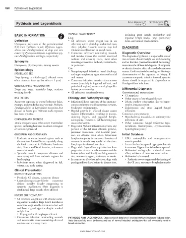
• Cutaneous infection: nonhealing wounds PYTHIOSIS AND LAGENIDIOSIS Cross-section of distal small intestinal Pythium insidiosum–induced lesion.
and invasive skin masses containing ulcerated Note characteristic mural thickening and loss of normal intestinal architecture that will eventually result in
nodules and draining tracts bowel obstruction.
www.ExpertConsult.com