
Page 1948 - Cote clinical veterinary advisor dogs and cats 4th
P. 1948
974 Thymoma
• Anorexia and weight loss also common • Thoracic ultrasound: morphologic exam of invasion into or attachment to local vessels.
• Weakness: associated with myasthenia or mass, including association with/invasion The size of thymoma is not necessarily predic-
VetBooks.ir • Regurgitation: if megaesophagus is present of cysts; fine-needle aspiration or needle ○ Chemotherapy: lymphoma protocol (pp.
hypercalcemia
of vascular structures, presence or absence
tive of complete or incomplete resection.
biopsy of mass (rule out lymphoma); col-
• Polyuria/polydipsia if hypercalcemic
607 and 609)
lection of pleural fluid (cytologic exam [pp.
• Dermatitis in cats (12%)
○ Radiation therapy
1164 and 1343]) • Nonresectable masses: radiation therapy ±
PHYSICAL EXAM FINDINGS • Survey abdominal radiographs, ultrasound: chemotherapy
• Respiratory system: muffled or absent lung rule out abdominal organ involvement • Myasthenia gravis (p. 668)
and heart sounds due to pleural effusion (lymphoma) ○ Resolution after complete resection of
and/or space-occupying nature of mass neoplasm
○ Increased, harsh lung sounds: aspiration Advanced or Confirmatory Testing ○ Prednisone 2 mg/kg PO q 24h for 4
pneumonia • MRI (p. 1132) or CT: determine whether weeks, then tapered
• Weakness: episodic or sustained; associated surgical excision is a viable treatment option. • Hypercalcemia (p. 491)
with loss of muscle tone, decreased spinal Evaluate compression, displacement, invasion, • Aspiration pneumonia (p. 793): antibiotic
reflexes. Can be focal, associated with the or incorporation of adjacent structures (e.g., therapy based on culture and susceptibility
pharyngeal, laryngeal, or facial muscles vessels, esophagus, trachea, thoracic wall) and (C&S) testing
• Decreased chest wall compliance is noted with metastasis. Evaluation of invasion of major • Megaesophagus (p. 642): consider tube
manual compression of the cranial thorax. vessels, including the cranial vena cava, is gastrostomy for medical and nutritional
○ Important physical finding in cats with difficult. Contrast administration does not support (p. 1109)
medium to large thymomas assist in determining cell type of mediastinal • Therapeutic thoracocentesis (p. 1164)
masses. Extent of mediastinal masses is often ○ If respiratory compromise due to pleural
Etiology and Pathophysiology unknown until surgery. effusion
• Neoplastic transformation of thymic epithe- • Acetylcholine (ACh) receptor antibody titers: ○ Preanesthetic patient stabilization
lial cells positive with thymoma-associated myasthenia
• Cranial mediastinal mass ± pleural effusion: gravis Possible Complications
clinical signs of respiratory compromise • Flow cytometry of samples collected by Inability to resolve the associated myasthenia
• Paraneoplastic syndrome of myasthenia gravis fine-needle aspiration and stored in fetal gravis:
result of aberrant immune stimulation bovine serum may be useful in differentiat- • Persistent regurgitation/megaesophagus
ing thymoma from lymphoma. The test • Recurring aspiration pneumonia
DIAGNOSIS uses T-cell marker expression of CD4 and • Intraoperative hemorrhage
CD8 on T cells; > 20% expression of CD4 • Disseminated intravascular coagulation
Diagnostic Overview and CD8 is consistent with thymoma, and • Perioperative mortality of 20% in dogs and
The diagnosis of thymoma is suspected based lack of CD4 and CD8 expression rules out 22% in cats due to nonresectable disease,
on presenting history, physical exam findings, thymoma. hemorrhage, or cardiopulmonary arrest
and demonstration of a well-defined cranial Regrowth of thymoma: occurs in some cases
mediastinal mass on thoracic radiographs. TREATMENT after complete surgical resection, as well as
Ultrasound-guided fine-needle aspirates or in cases with incomplete surgical resection.
needle biopsies are required to confirm the Treatment Overview Monitoring with exam and imaging studies
diagnosis and rule out other causes such as Complete surgical resection is the recommended is essential regardless of completeness of
mediastinal lymphoma. Computerized tomog- treatment. resection.
raphy (CT) helps determine whether the mass
is surgically resectable. Acute and Chronic Treatment Recommended Monitoring
• Complete surgical excision is often feasible • After surgical resection (complete or
Differential Diagnosis with thymoma. May be carried out thora- incomplete): q 3 months (exam, thoracic
• Thymic lymphoma, hyperplasia, or hemorrhage coscopically for noninvasive thymoma radiographs) for 1 year, then q 6 months
• Branchial cyst • Incomplete surgical excision is more likely • If patient is receiving chemotherapy: as
• Ectopic thyroid or parathyroid neoplasia when thoracic ultrasonography reveals determined by protocol
• Neurogenic tumor
• Heart base tumor
• Mediastinal abscess or granuloma
Initial Database
• CBC, serum biochemical profile, and urinalysis:
evaluate for hypercalcemia (5%-35% of dogs
hypercalcemic), which does not change survival
time; poorly concentrated urine if hypercal-
cemia present; inflammatory leukogram if
aspiration pneumonia due to megaesophagus
• Feline leukemia virus (FeLV) and feline
immunodeficiency (FIV) serologic testing
in cats: thymic lymphoma often associated
with FeLV-positive status
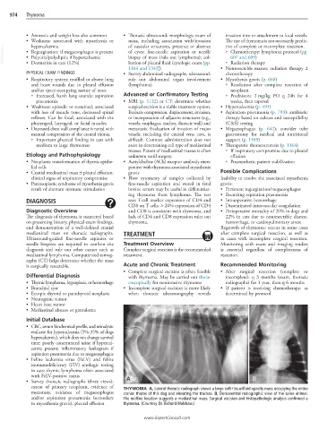
• Survey thoracic radiographs (three views): A B
extent of primary neoplasm, evidence of THYMOMA A, Lateral thoracic radiograph shows a large soft-tissue/fluid opacity mass occupying the entire
metastasis, evidence of megaesophagus cranial thorax of this dog and elevating the trachea. B, Dorsoventral radiographic view of the same animal.
and/or aspiration pneumonia (secondary The midline location suggests a mediastinal mass. Surgical excision and histopathologic analysis confirmed a
to myasthenia gravis), pleural effusion thymoma. (Courtesy Dr. Richard Walshaw.)
www.ExpertConsult.com