
Page 2225 - Cote clinical veterinary advisor dogs and cats 4th
P. 2225
Feeding Tube Placement: Esophagostomy 1107.e1
VetBooks.ir
Internal
jugular vein
Internal
jugular vein Common
carotid artery
Common
carotid artery Procedures and Techniques
Forceps
Esophagus
External
Trachea Esophagus jugular vein Trachea External
jugular vein
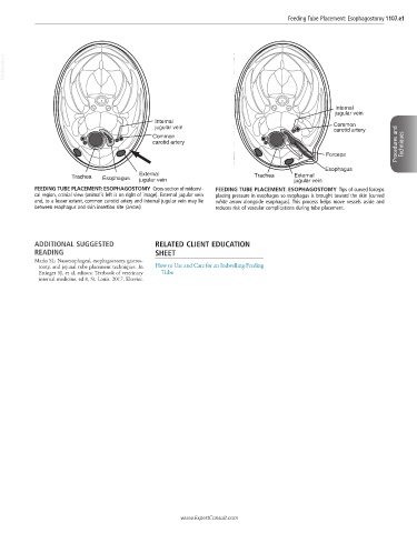
FEEDING TUBE PLACEMENT: ESOPHAGOSTOMY Cross-section of midcervi- FEEDING TUBE PLACEMENT: ESOPHAGOSTOMY Tips of curved forceps
cal region, cranial view (animal’s left is on right of image). External jugular vein placing pressure in esophagus so esophagus is brought toward the skin (curved
and, to a lesser extent, common carotid artery and internal jugular vein may lie white arrow alongside esophagus). This process helps move vessels aside and
between esophagus and skin insertion site (arrow). reduces risk of vascular complications during tube placement.
ADDITIONAL SUGGESTED RELATED CLIENT EDUCATION
READING SHEET
Marks SL: Nasoesophageal, esophagostomy, gastros-
tomy, and jejunal tube placement techniques. In How to Use and Care for an Indwelling Feeding
Ettinger SJ, et al, editors: Textbook of veterinary Tube
internal medicine, ed 8, St. Louis. 2017, Elsevier.
www.ExpertConsult.com