
Page 226 - Cote clinical veterinary advisor dogs and cats 4th
P. 226
Atrial Premature Complexes and Atrial Tachycardia 97
which may be reversible with appropriate Vagal maneuver: ○ Intravenous (IV) treatment if extremely
treatment. • May suddenly terminate atrial and junctional rapid rate and/or causing clinical signs
VetBooks.ir DIAGNOSIS • Should have no effect on ventricular ○ Oral medications to reduce long-term Diseases and Disorders
(e.g., severe anxiety, syncope)
tachycardias or slow rate to aid in diagnosis
effect of uncontrolled tachycardia on
tachycardia
Diagnostic Overview
may be used in diagnosing underlying cardiac
Initial suspicion is usually based on auscultation Thoracic radiography and echocardiography: myocardial function
of premature beats or a rapid heart rate. Defini- disease or concurrent CHF. Acute General Treatment
tive diagnosis is made based on the electrocar- • AT/SVT producing sustained ventricular
diogram (ECG). Advanced or Confirmatory Testing rates > 250 beats/min in dogs often severely
Electrophysiologic studies: may be used for compromises diastolic ventricular filling and
Differential Diagnosis determining the underlying mechanism of can be considered critical; IV antiarrhythmics
• AT must be differentiated from sinus arrhythmia. Not widely available. to decrease ventricular rate are warranted.
tachycardia; sinus tachycardia is usually an Patients should have an IV catheter and
appropriate physiologic tachycardia in TREATMENT continuous ECG monitoring during drug
response to pain or anxiety. administration.
• Uncommonly, atrial arrhythmias may coexist Treatment Overview • Perform vagal maneuver first. Some ATs/
with aberrant conduction/bundle branch • Always correct underlying cause or contribut- SVTs terminate with ocular pressure or
block, producing wide QRS complexes and ing factors first (e.g., CHF/hypoxemia, carotid sinus massage. If not, judicious IV
causing APCs and AT to appear similar to hypokalemia, acidosis, hypovolemia). drugs are warranted.
VPCs and ventricular tachycardia, respectively. • Return hemodynamic stability, especially with • Calcium channel blockers: commonly used
With APCs and AT/SVT, a P′ wave precedes continuous rapid AT; isolated, infrequent as first-choice agents:
each QRS complex at a repeatable interval APCs do not cause hemodynamic instability, ○ Diltiazem: 0.05 mg/kg IV over 1-2 min,
(sometimes buried in preceding T wave). and no specific treatment is required. repeat prn to total dose of 0.75 mg/kg,
• Conversion of the arrhythmia to sinus or
Initial Database rhythm is not always possible, especially with ○ Verapamil: 0.05 mg/kg IV q 5-10 min
ECG (APCs): atrial enlargement (substrate for arrhythmia up to total dose of 0.15 mg/kg, or 2-10
• A P′ wave represents premature atrial recurrence/persistence). micrograms/kg/min constant rate infusion
depolarization. Its ectopic origin (outside the • Control the ventricular response rate if too • Beta-blockers:
sinoatrial node) means it propagates differ- rapid. ○ Esmolol: 0.05-0.5 mg/kg slow IV; can
ently through the atria than a sinus-origin ○ Target rate is achieved with drugs that follow with 0.05-0.1 mg/kg/min constant
impulse, and P′ waves are therefore of dif- slow AV node conduction to optimize the rate infusion if needed, or
ferent shape and occur sooner (prematurely) ventricular rate. ○ Propranolol: 0.02 mg/kg IV slowly over
compared to the expected normal P waves. ○ Target rate varies with the underlying 2-3 minutes, titrate dose up to effect (to
• The complete heartbeat (P′-QRS-T) occurs cardiac disease. maximum of 0.1 mg/kg)
earlier than the next expected sinus beat.
• P′ waves may not be visible if the rate is so
fast that they are buried in the preceding T Z
wave or they are isoelectric in that lead * *
(examine other ECG leads).
• QRS complexes typically are narrow and
positive in lead II, like the patient’s sinus
QRS complexes.
• APCs are usually followed by a noncompen-
satory pause; the ectopic atrial impulse resets
the sinus node, such that the R-R interval of
two normal sinus complexes enclosing the
APC is less than the R-R intervals of three A
consecutive sinus complexes.
ECG (AT):
• Three or more APCs in a row; regular or
slightly irregular rhythm I I
• P′ waves are present but may be hidden or
superimposed on preceding T waves.
• The onset and termination at AT is usually
sudden (paroxysmal) and does not speed up
or slow down.
• The P′R interval is usually constant.
• Narrow QRS complexes (rarely, can be wide
with coexisting bundle branch block or
aberrant conduction)
• At extremely rapid atrial rates, there may be B
varying degrees of AV block (i.e., noncon-
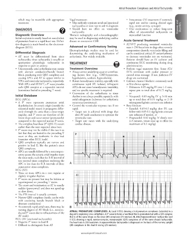
ducted P′ waves due to refractoriness of the ATRIAL PREMATURE COMPLEXES A, Lead II ECG showing atrial premature complexes (asterisks) in a
dog with respiratory sinus arrhythmia. A P′ wave initiates a heartbeat that is premature but with a QRS complex
AV node). that is of the same shape as the sinus QRS complexes (25 mm/sec). B, Atrial/supraventricular tachycardia lead
ECG (AV junctional tachycardia): II ECG showing a rapid, narrow-complex, monomorphic (QRS complexes all of the same shape) tachycardia.
• Negative P′ waves in lead II The heart rate is 330 beats/min. A supraventricular tachycardia is diagnosed on the basis of the narrow, upright
• Difficult to distinguish from AT QRS complexes in this lead II tracing (25 mm/sec).
www.ExpertConsult.com