
Page 2260 - Cote clinical veterinary advisor dogs and cats 4th
P. 2260
Intravenous Catheter Placement 1123.e5
VetBooks.ir
Procedures and Techniques
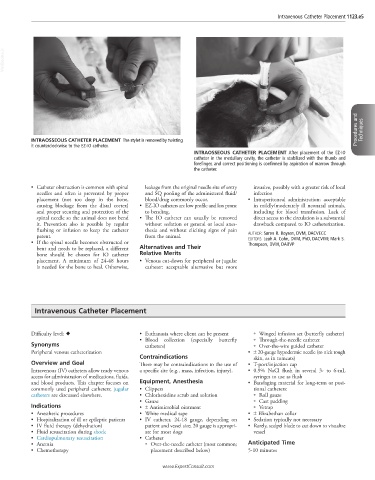
INTRAOSSEOUS CATHETER PLACEMENT The stylet is removed by twisting
it counterclockwise to the EZ-IO catheter.
INTRAOSSEOUS CATHETER PLACEMENT After placement of the EZ-IO
catheter in the medullary cavity, the catheter is stabilized with the thumb and
forefinger, and correct positioning is confirmed by aspiration of marrow through
the catheter.
• Catheter obstruction is common with spinal leakage from the original needle site of entry invasive, possibly with a greater risk of local
needles and often is prevented by proper and SQ pooling of the administered fluid/ infection
placement (not too deep in the bone, blood/drug commonly occur. • Intraperitoneal administration: acceptable
causing blockage from the distal cortex) • EZ-IO catheters are low profile and less prone in mildly/moderately ill neonatal animals,
and proper securing and protection of the to bending. including for blood transfusion. Lack of
spinal needle so the animal does not bend • The IO catheter can usually be removed direct access to the circulation is a substantial
it. Prevention also is possible by regular without sedation or general or local anes- drawback compared to IO catheterization.
flushing or infusion to keep the catheter thesia and without eliciting signs of pain
patent. from the animal. AUTHOR: Søren R. Boysen, DVM, DACVECC
• If the spinal needle becomes obstructed or EDITORS: Leah A. Cohn, DVM, PhD, DACVIM; Mark S.
Thompson, DVM, DABVP
bent and needs to be replaced, a different Alternatives and Their
bone should be chosen for IO catheter Relative Merits
placement. A minimum of 24-48 hours • Venous cut-down for peripheral or jugular
is needed for the bone to heal. Otherwise, catheter: acceptable alternative but more
Intravenous Catheter Placement
Difficulty level: ♦ • Euthanasia where client can be present ○ Winged infusion set (butterfly catheter)
• Blood collection (especially butterfly ○ Through-the-needle catheter
Synonyms catheters) ○ Over-the-wire guided catheter
Peripheral venous catheterization • ± 20-gauge hypodermic needle (to nick tough
Contraindications skin, as in tomcats)
Overview and Goal There may be contraindications to the use of • T-port/injection cap
Intravenous (IV) catheters allow ready venous a specific site (e.g., mass, infection, injury). • 0.9% NaCl flush in several 3- to 6-mL
access for administration of medications, fluids, syringes to use as flush
and blood products. This chapter focuses on Equipment, Anesthesia • Bandaging material for long-term or posi-
commonly used peripheral catheters; jugular • Clippers tional catheters:
catheters are discussed elsewhere. • Chlorhexidine scrub and solution ○ Roll gauze
• Gauze ○ Cast padding
Indications • ± Antimicrobial ointment ○ Vetrap
• Anesthetic procedures • White medical tape • ± Elizabethan collar
• Hospitalization of ill or epileptic patients • IV catheter, 24-18 gauge, depending on • Sedation typically not necessary
• IV fluid therapy (dehydration) patient and vessel size; 20 gauge is appropri- • Rarely, scalpel blade to cut down to visualize
• Fluid resuscitation during shock ate for most dogs vessel
• Cardiopulmonary resuscitation • Catheter
• Anemia ○ Over-the-needle catheter (most common; Anticipated Time
• Chemotherapy placement described below) 5-10 minutes
www.ExpertConsult.com