
Page 232 - Cote clinical veterinary advisor dogs and cats 4th
P. 232
Atrial Septal Defect 99.e3
VetBooks.ir RV RV Diseases and Disorders
RA RA
LV HR=138bpm
LV
LA
LA
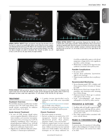
ATRIAL SEPTAL DEFECT Right, parasternal, long-axis, four-chamber view of
ATRIAL SEPTAL DEFECT Right, parasternal, long-axis, four-chamber view of a patient with a small septum secundum atrial septal defect after an intravenous
the heart in a dog with an atrial septal defect (arrow). Note the distinct margins injection of agitated saline. Note the presence of multiple echo artifacts from saline
and relatively dorsal positioning of the defect in the interatrial septum, which helps microbubbles in all four cardiac chambers (arrows), consistent with right-to-left
distinguish this lesion from the fossa ovalis and normal echo dropout. Color flow shunting at the atrial level. LA, Left atrium; LV, left ventricle; RA, right atrium; RV,
and spectral Doppler are useful to document flow through the defect. LA, Left right ventricle.
atrium; LV, left ventricle; RA, right atrium; RV, right ventricle.
should be considered for patients with clinical
symptoms or larger defects with significant
secondary cardiac changes.
• Phlebotomy as indicated for hyperviscosity
syndrome associated with erythrocytosis
Possible Complications
RA HR=150bpm • Recurrent CHF
LV • Syncope (from pulmonary hypertension,
right-to-left shunting)
• Hyperviscosity syndrome due to erythrocy-
tosis
LA
Recommended Monitoring
• Serum chemistry panel, CBC, urinalysis, and
blood pressure before initiation of therapy
for CHF
FOSSA OVALIS Right, parasternal, long-axis, four-chamber view of a normal dog with echo dropout in the • Blood pressure, blood urea nitrogen (BUN),
region of the fossa ovalis. Note the indistinct margins (arrow) and tapering septal echoes, both of which help creatinine, and electrolytes after initiating
distinguish this from a small atrial septal defect. LA, Left atrium; LV, left ventricle; RA, right atrium. heart failure therapy
• Hematocrit/arterial blood gas in patients
with right-to-left shunting
TREATMENT • A positive inotrope (pimobendan) may be • Serial echocardiography and thoracic
indicated in cases with severe myocardial radiographs as dictated by defect severity
Treatment Overview failure.
Prevent/delay increases in pulmonary vascular • Abdominocentesis/thoracocentesis as neces- PROGNOSIS & OUTCOME
resistance and CHF. With larger defects, this sary for body cavity effusions
should include consideration for definitive • Phlebotomy as indicated for hyperviscosity • Excellent with mild (small) congenital defects
repair and closure of the defect. syndrome associated with erythrocytosis • Guarded to poor with large defects, pulmo-
nary hypertension, right-to-left shunting, or
Acute General Treatment Chronic Treatment animals with CHF
• In cases of CHF, therapy should reduce • Recurrent centesis as necessary • Poor with acquired defects associated with
venous congestion (diuretics), inhibit sodium/ • Recheck evaluations are essential for CHF severe valvular disease
water retention, and counteract vasoconstric- management (monitor renal function,
tion (angiotensin-converting enzyme [ACE] albumin/total protein, blood pressure, PEARLS & CONSIDERATIONS
inhibitors, vasodilators). Type 5 and type 3 electrolyte balance, heart rate/rhythm).
phosphodiesterase inhibitors (e.g., sildenafil • Definitive surgical repair has been reported Comments
and pimobendan, respectively) may be useful and is available at selected academic institu- A common error of inexperienced echocardiog-
with severe pulmonary hypertension. tions. Catheter closure has also been reported raphers is the misdiagnosis of normal, mid-atrial
• Digoxin may be indicated with atrial tachyar- and is more widely available. These advanced septal echo dropout (the normal fossa ovalis)
rhythmias or baroreceptor dysfunction. techniques are not always indicated but as an ASD.
www.ExpertConsult.com