
Page 2354 - Cote clinical veterinary advisor dogs and cats 4th
P. 2354
Thoracocentesis 1165
Possible Complications and
Common Errors to Avoid
VetBooks.ir • Intrathoracic hemorrhage
• Iatrogenic pneumothorax
• Re-expansion pulmonary edema in situations
of chronic pleural space disease
• Acute death from stress of restraint in animals
with severe respiratory compromise A
Procedure
• Position animal, preferably in sternal recum-
bency or standing, but lateral recumbency
is also acceptable for pneumothorax. D E
• Provide supplemental oxygen if needed
(p. 1146).
• Have assistant available to restrain animal C Procedures and Techniques
or give sedation as needed.
○ Consider brief, quiet rest in oxygen B
cage if patient is anxious and extremely
dyspneic.
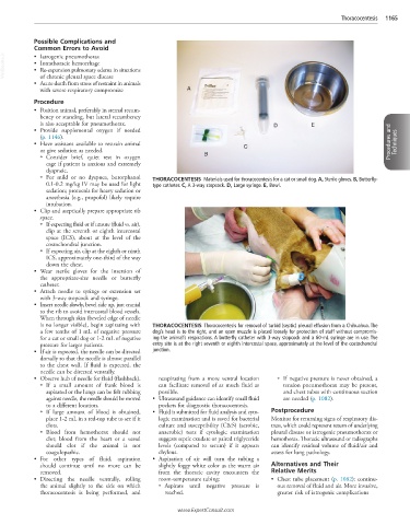
○ For mild or no dyspnea, butorphanol THORACOCENTESIS Materials used for thoracocentesis for a cat or small dog. A, Sterile gloves. B, Butterfly-
0.1-0.2 mg/kg IV may be used for light type catheter. C, A 3-way stopcock. D, Large syringe. E, Bowl.
sedation; protocols for heavy sedation or
anesthesia (e.g., propofol) likely require
intubation.
• Clip and aseptically prepare appropriate rib
space.
○ If expecting fluid or if unsure (fluid vs. air),
clip at the seventh or eighth intercostal
space (ICS), about at the level of the
costochondral junction.
○ If expecting air, clip at the eighth or ninth
ICS, approximately one-third of the way
down the chest.
• Wear sterile gloves for the insertion of
the appropriate-size needle or butterfly
catheter.
• Attach needle to syringe or extension set
with 3-way stopcock and syringe.
• Insert needle slowly, bevel side up, just cranial
to the rib to avoid intercostal blood vessels.
When through skin (beveled edge of needle
is no longer visible), begin aspirating with THORACOCENTESIS Thoracocentesis for removal of turbid (septic) pleural effusion from a Chihuahua. The
a few tenths of 1 mL of negative pressure dog’s head is to the right, and an open muzzle is placed loosely for protection of staff without compromis-
for a cat or small dog or 1-2 mL of negative ing the animal’s respirations. A butterfly catheter with 3-way stopcock and a 60-mL syringe are in use. The
pressure for larger patients. entry site is at the right seventh or eighth intercostal space, approximately at the level of the costochondral
• If air is expected, the needle can be directed junction.
dorsally so that the needle is almost parallel
to the chest wall. If fluid is expected, the
needle can be directed ventrally.
• Observe hub of needle for fluid (flashback). reaspirating from a more ventral location ○ If negative pressure is never obtained, a
○ If a small amount of frank blood is can facilitate removal of as much fluid as tension pneumothorax may be present,
aspirated or the lungs can be felt rubbing possible. and chest tubes with continuous suction
against needle, the needle should be moved • Ultrasound guidance can identify small fluid are needed (p. 1082).
to a different location. pockets for diagnostic thoracocentesis.
○ If large amount of blood is obtained, • Fluid is submitted for fluid analysis and cyto- Postprocedure
place 1-2 mL in a red-top tube to see if it logic examination and is saved for bacterial Monitor for returning signs of respiratory dis-
clots. culture and susceptibility (C&S) (aerobic, tress, which could represent return of underlying
○ Blood from hemothorax should not anaerobic) tests if cytologic examination pleural disease or iatrogenic pneumothorax or
clot; blood from the heart or a vessel suggests septic exudate or paired triglyceride hemothorax. Thoracic ultrasound or radiographs
should clot if the animal is not levels (compared to serum) if it appears can identify residual volume of fluid/air and
coagulopathic. chylous. assess for lung pathology.
• For other types of fluid, aspiration • Aspiration of air will turn the tubing a
should continue until no more can be slightly foggy white color as the warm air Alternatives and Their
removed. from the thoracic cavity encounters the Relative Merits
• Directing the needle ventrally, rolling room-temperature tubing: • Chest tube placement (p. 1082): continu-
the animal slightly to the side on which ○ Aspirate until negative pressure is ous removal of fluid and air. More invasive,
thoracocentesis is being performed, and reached. greater risk of iatrogenic complications
www.ExpertConsult.com