
Page 88 - Cote clinical veterinary advisor dogs and cats 4th
P. 88
Actinomycosis and Nocardiosis 21.e1
VetBooks.ir Diseases and Disorders
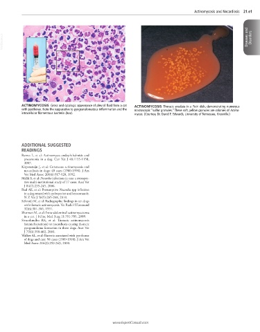
ACTINOMYCOSIS Gross and cytologic appearance of pleural fluid from a cat ACTINOMYCOSIS Thoracic exudate in a Petri dish, demonstrating numerous
with pyothorax. Note the suppurative to pyogranulomatous inflammation and the macroscopic “sulfur granules.” These soft, yellow granules are colonies of Actino-
intracellular filamentous bacteria (box). myces. (Courtesy Dr. David F. Edwards, University of Tennessee, Knoxville.)
ADDITIONAL SUGGESTED
READINGS
Barnes L, et al: Actinomyces endophthalmitis and
pneumonia in a dog. Can Vet J 48:1155-1158,
2007.
Kirpensteijn J, et al: Cutaneous actinomycosis and
nocardiosis in dogs: 48 cases (1980-1990). J Am
Vet Med Assoc 201(6):917-920, 1992.
Malik R, et al: Nocardia infections in cats: a retrospec-
tive multi-institutional study of 17 cases. Aust Vet
J 84(7):235-245, 2006.
Paul AE, et al: Presumptive Nocardia spp infection
in a dog treated with cyclosporine and ketoconazole.
N Z Vet J 58(5):265-268, 2010.
Schmidt M, et al: Radiographic findings in ten dogs
with thoracic actinomycosis. Vet Radiol Ultrasound
32(6):301-306, 1991.
Sharman M, et al: Intra-abdominal actinomycetoma
in a cat. J Feline Med Surg 11:701-705, 2009.
Sivacolundhu RK, et al: Thoracic actinomycosis
(arcanobacteriosis) or nocardiosis causing thoracic
pyogranuloma formation in three dogs. Aust Vet
J 79(6):398-402, 2001.
Walker AL, et al: Bacteria associated with pyothorax
of dogs and cats: 98 cases (1989-1998). J Am Vet
Med Assoc 216(3):359-363, 2000.
www.ExpertConsult.com