
Page 954 - Cote clinical veterinary advisor dogs and cats 4th
P. 954
Hip Dysplasia 469
PROGNOSIS & OUTCOME
VetBooks.ir • Good prognosis Diseases and Disorders
○ If the patient has no or mild clinical signs
○ With surgical intervention in the absence
of aspiration pneumonia
• Guarded to poor prognosis associated with
○ Severe aspiration pneumonia
○ Severe esophagitis/stricture
○ Persistent megaesophagus
PEARLS & CONSIDERATIONS
Comments
• Consider referral to a soft-tissue surgeon if
surgical correction is indicated.
• Treatment for esophagitis should be
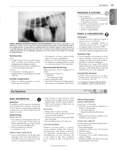
HIATAL HERNIA/GASTROESOPHAGEAL INTUSSUSCEPTION Lateral thoracic radiograph of a dog
demonstrates a large, soft-tissue mass due to gastroesophageal intussusception, located characteristically in continued postoperatively for any patient
the caudodorsal thorax. The dog suffered strangulation injury when it became caught in a snare. Also visible is undergoing surgical correction of HH.
an air-filled esophagus, which demonstrates both sides of the dorsal tracheal membrane (tracheal stripe sign). • Aspiration pneumonia requires intensive treat-
A differential diagnosis for the radiographic appearance of the mass lesion is primary lung tumor. (Courtesy ment before considering surgical intervention.
Dr. Richard Walshaw.)
Technician Tips
Nutrition/Diet • Overreduction of hiatal opening during • Pressure applied to the abdomen via a belly
Acute: surgery may worsen regurgitation. wrap or pressing a wooden spoon on the
• Upright feeding of low-fat, small, frequent • Recurrence of HH after surgical correction abdomen can improve the chance of observ-
meals to promote normal function of the • Development of esophageal stricture second- ing HH on abdominal radiographs.
esophagus/gastroesophageal junction ary to gastroesophageal reflux/esophagitis • Positioning on the surgery table with the
• Feeding tube (percutaneous endogastric [p. abdomen lower than the head allows abdomi-
1109]) if oral intake inadequate Recommended Monitoring nal organs to fall caudally, thereby improving
Chronic: Follow-up positive contrast esophagram (4-6 visibility and access to the diaphragm.
• Continuation of feeding regimen weeks):
• Upright feeding if megaesophagus associated • Demonstrate resolution of SUGGESTED READING
with GEI ○ HH/GEI Cornell KL: Stomach. In Johnston SA, et al., editors:
○ Gastroesophageal reflux Veterinary surgery: small animal, ed 2, St. Louis,
Possible Complications • Assess esophageal motility. 2017, Elsevier pp 1700-1730.
• Failure of medical management to resolve • Determine if continuation of feeding regimen AUTHOR: MaryAnn G. Radlinsky, DVM, MS, DACVS
clinical signs of esophagitis and treatment of esophagitis still necessary EDITOR: Elizabeth A. Swanson, DVM, MS, DACVS
Video
Hip Dysplasia Client Education Available
Sheet
BASIC INFORMATION • Most common in large and giant breeds Clinical Presentation
(Labrador retriever, golden retriever, German
Definition shepherd dog, rottweiler, Newfoundland) DISEASE FORMS/SUBTYPES
A condition caused by abnormal development • May be more common in Maine coon cats Juvenile (up to 18 months):
of the coxofemoral (i.e., hip) joint, characterized than other cat breeds • Hip laxity characterized by subluxation or
by joint laxity in young patients progressing luxation of the hip joint
to degenerative joint disease (DJD) of various RISK FACTORS ○ Acetabular dysplasia
degrees of severity • Being a large- or giant-breed dog ○ Femoral head/neck malformations
• Castration of young male dogs may promote Mature:
Epidemiology expression of hip dysplasia. • Degenerative joint disease secondary to hip
SPECIES, AGE, SEX • Excessive caloric intake causing rapid weight instability and incongruity
• The condition is a developmental disease and gain and growth
occurs in young dogs. Clinical signs may • Synovial inflammation of the hip joint HISTORY, CHIEF COMPLAINT
become apparent at any age, or the condition Juvenile patients typically have hindlimb lame-
may remain occult throughout life. ASSOCIATED DISORDERS ness characterized by
• No sex predisposition reported Common concurrent genetic or develop- • Bunny-hopping gait
• Less frequent in cats mental conditions include elbow dysplasia, • Unilateral or bilateral pelvic limb lameness
osteochondrosis, stifle disease, panosteitis, • Difficulty rising
GENETICS, BREED PREDISPOSITION and hypertrophic osteodystrophy. A causal • Exercise intolerance
• Cause is not fully known, but the phenotypic relationship has not been identified, and • Description of an audible clicking when
expression of hip laxity that progresses to it may simply be that these conditions rising or walking
early DJD is due to the patient’s genotype are all common in large- and giant-breed • Shifting of weight to the thoracic limbs and
as well as environmental influences. dogs. hyperextension of the hocks
www.ExpertConsult.com