
Page 125 - A Practical Guide to Equine Radiography
P. 125
106 A PRACTICAL GUIDE TO EQUINE RADIOGRAPHY
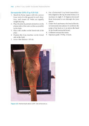
Dorsoplantar (DPl) (Figs 11.5–11.8) 6. Use a horizontal X-ray beam perpendicu-
VetBooks.ir 1. Stand the horse square with the cannon larly aligned to the leg. In some horses, it is
necessary to angle 5–10 degrees downward
bone vertical to the ground in each direc-
tion, and ensure all limbs are equally from horizontal to see through the joint
weight-bearing. spaces.
2. Place the plate in portrait orientation on the 7. Centre the X-ray beam at the level of the dis-
plantar side of the joint as close as possible tal intertarsal joint (about 10 cm below the
to the limb. point of the hock or 2 cm above the head
3. Place a R/L marker on the lateral side of the of the fourth metatarsal bone).
plate. 8. Collimate around the tarsus.
4. Position the X-ray machine on the dorsal 9. Exposure guide: 70 kVp, 10 mAs.
side of the limb.
5. Focus–film distance: 100 cm.
Figure 11.5 Positioning to obtain a DPI view of the tarsus.
Equine Radiography.indb 106 27/11/2018 11:09