
Page 801 - Withrow and MacEwen's Small Animal Clinical Oncology, 6th Edition
P. 801
CHAPTER 34 Miscellaneous Tumors 779
Pathology and Natural Behavior
VetBooks.ir Thymomas are neoplasms of thymic epithelial cells, but they
commonly include other cell populations such as mast cells and
Different histologic types of thy-
136,139–141
mature lymphocytes.
moma have been described, including epithelial, lymphocyte-
rich, and clear cell. In cats, cystic thymomas seem to be the most
common form, but squamous cell carcinomas and thymolipoma
have also been reported. 136,140–144 Thymomas are carcinomas and
thus should be considered malignant tumors. The terms benign
or malignant thymoma are commonly used and are based on
clinical evidence of invasiveness rather than on histologic features
of malignancy. Metastasis is rare in both species, 141,145–147 but
reported metastatic rate has been as high as 20% in cats with cys-
tic thymomas. 142 The differential diagnoses for mediastinal masses
include lymphoma, ectopic thyroid tumor, branchial cysts, and,
rarely, sarcomas and metastatic neoplasms. It is important to note
that tumors extending from the ribs or sternum into the cranial
mediastinum may sometimes resemble a mediastinal mass. 148
History and Clinical Signs
Clinical signs related to organ displacement due to the pres-
ence of a mediastinal mass include lethargy, regurgitation, vom-
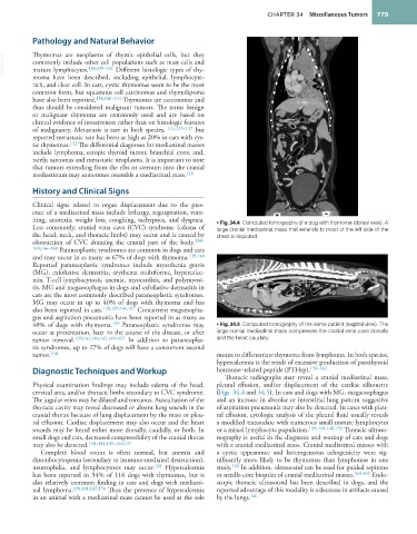
iting, anorexia, weight loss, coughing, tachypnea, and dyspnea. • Fig. 34.4 Computed tomography of a dog with thymoma (dorsal view). A
Less commonly, cranial vena cava (CVC) syndrome (edema of large cranial mediastinal mass that extends to most of the left side of the
the head, neck, and thoracic limbs) may occur and is caused by chest is depicted.
obstruction of CVC draining the cranial part of the body. 138–
143,146–148 Paraneoplastic syndromes are common in dogs and cats
and may occur in as many as 67% of dogs with thymoma. 139,140
Reported paraneoplastic syndromes include myasthenia gravis
(MG), exfoliative dermatitis, erythema multiforme, hypercalce-
mia, T-cell lymphocytosis, anemia, myocarditis, and polymyosi-
tis. MG and megaesophagus in dogs and exfoliative dermatitis in
cats are the most commonly described paraneoplastic syndromes.
MG may occur in up to 40% of dogs with thymoma and has
also been reported in cats. 138,139,146,147 Concurrent megaesopha-
gus and aspiration pneumonia have been reported in as many as
40% of dogs with thymoma. 139 Paraneoplastic syndromes may • Fig. 34.5 Computed tomography of the same patient (sagittal view). The
occur at presentation, later in the course of the disease, or after large cranial mediastinal mass compresses the cranial vena cava dorsally
tumor removal. 139,141,146,147,149–157 In addition to paraneoplas- and the heart caudally.
tic syndromes, up to 27% of dogs will have a concurrent second
tumor. 138 means to differentiate thymoma from lymphoma. In both species,
hypercalcemia is the result of excessive production of parathyroid
Diagnostic Techniques and Workup hormone–related peptide (PTHrp). 158–162
Thoracic radiographs may reveal a cranial mediastinal mass,
Physical examination findings may include edema of the head, pleural effusion, and/or displacement of the cardiac silhouette
cervical area, and/or thoracic limbs secondary to CVC syndrome. (Figs. 34.4 and 34.5). In cats and dogs with MG, megaesophagus
The jugular veins may be dilated and tortuous. Auscultation of the and an increase in alveolar or interstitial lung pattern suggestive
thoracic cavity may reveal decreased or absent lung sounds in the of aspiration pneumonia may also be detected. In cases with pleu-
cranial thorax because of lung displacement by the mass or pleu- ral effusion, cytologic analysis of the pleural fluid usually reveals
ral effusion. Cardiac displacement may also occur and the heart a modified transudate with numerous small mature lymphocytes
sounds may be heard either more dorsally, caudally, or both. In or a mixed lymphocyte population. 139–141,146,159 Thoracic ultraso-
small dogs and cats, decreased compressibility of the cranial thorax nography is useful in the diagnosis and workup of cats and dogs
may also be detected. 138–141,145–146,157 with a cranial mediastinal mass. Cranial mediastinal masses with
Complete blood count is often normal, but anemia and a cystic appearance and heterogeneous echogenicity were sig-
thrombocytopenia (secondary to immune-mediated destruction), nificantly more likely to be thymomas than lymphomas in one
neutrophilia, and lymphocytosis may occur. 138 Hypercalcemia study. 163 In addition, ultrasound can be used for guided aspirates
has been reported in 34% of 116 dogs with thymomas, but is or needle-core biopsies of cranial mediastinal masses. 164,165 Endo-
also relatively common finding in cats and dogs with mediasti- scopic thoracic ultrasound has been described in dogs, and the
nal lymphoma. 138,141,147,158 Thus the presence of hypercalcemia reported advantage of this modality is a decrease in artifacts caused
in an animal with a mediastinal mass cannot be used as the sole by the lungs. 166