
Page 1004 - Small Animal Internal Medicine, 6th Edition
P. 1004
976 PART VIII Reproductive System Disorders
calciuria, decrease intestinal calcium absorption, and impair (periurethral) vaginal mucosa to estrogen, can recur near
osteoclasia. Hypoglycemia should be corrected if present, parturition and can be differentiated by physical examina-
VetBooks.ir and exogenous treatment for hyperthermia given if tion or vaginoscopy (see Fig. 55.5, B).
Rupture of the uterus occurs most commonly with very
necessary.
Once the immediate neurologic signs are controlled, a SC
uterine wall, especially in multiparous dams with dystocia.
injection of the equal volume of calcium gluconate, diluted large litters causing marked stretching and thinning of the
50% with saline is given and repeated q6-8h until the dam Immediate laparotomy for retrieval of fetuses and repair or
is stable and able to take oral supplementation. Calcium removal of the uterus, as well as culture and lavage of the
gluconate or carbonate (10-30 mg/kg q8h) should be insti- abdominal cavity, is indicated. The uterus should be carefully
tuted. Each 500-mg calcium carbonate tablet (Tums) sup- examined at any cesarean section for any areas with or prone
plies 200 mg calcium. Efforts to diminish lactational to rupture. Peritonitis or hemoabdomen can result from an
demands on the dam and improve her plane of nutrition are undetected uterine tear. A unilateral hysterectomy can be
indicated. If response to therapy has been prompt, nursing considered if the damaged area is limited and the dam valu-
can be gradually reinstituted until the neonates can be safely able to a breeding program; these females can be prone to
weaned, usually at a slightly early age (3 weeks), and concur- uterine torsion as a consequence.
rent supplementation with commercial bitch/queen milk
replacement is encouraged. Bottle feeding should precede Subinvolution of Placental Sites
nursing, and nursing limited to 10 min q4-6h rather than The persistence of serosanguineous to hemorrhagic vaginal
continuously. Giving calcium throughout lactation (but not discharge beyond 16 weeks postpartum can indicate subin-
prepartum) may be attempted in dams with a history of volution of the placental sites of attachment (SIPS) in the
recurrent eclampsia (calcium carbonate 500-4000 mg/dam/ bitch. Histologically, fetal trophoblastic cells have persisted
day, divided). in the myometrium instead of degenerating, endometrial
vessel thrombosis is lacking, and normal involution of the
uterus is prevented. Normal interplacental regions exist.
UTERINE DISORDERS Eosinophilic masses of collagen and dilated endometrial
Uterine Trauma glands protrude into the uterine lumen, oozing blood (Fig.
Complete or partial prolapse of the uterus is an uncom- 55.27). The cause is unknown, blood loss is usually minimal,
mon postpartum condition in the bitch, occurring rarely intrauterine infection not present, and fertility is unaffected.
in the queen experiencing dystocia. The diagnosis is based Treatment is generally not necessary; recovery is spontane-
on palpation of a firm, tubular mass protruding from the ous and symptoms mild. In the uncommon situation where
vulva postpartum, and inability to identify the uterus with vaginal bleeding from SIPS is copious enough to cause
abdominal ultrasonography. The prolapsed uterine tissues serious anemia, coagulopathies (likely defects in the intrinsic
are at risk for maceration and infection from exposure and pathway or thrombocytopenia/thrombocytopathies), uterine
contamination (Fig. 55.26). The size of most bitches and trauma, neoplasia of the genitourinary tract, severe metritis,
queens precludes manual replacement; laparotomy and and excessive hemorrhage from prematurely separated pla-
ovariohysterectomy are usually indicated. Vaginal hyperpla- cental sites should be ruled out. Cytology of vulvar discharge
sia and prolapse, secondary to a hypersensitivity of focal (trophoblast cells can be evident), vaginoscopy to localize
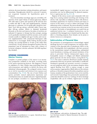
FIG 55.27
FIG 55.26 Gross specimen, subinvolution of placental sites. The right
Uterine prolapse postdystocia in a queen. Devitalized tissue horn has been incised to illustrate hematoma formation at
necessitated ovariohysterectomy. placental sites.