
Page 109 - Small Animal Internal Medicine, 6th Edition
P. 109
CHAPTER 4 Cardiac Arrhythmias and Antiarrhythmic Therapy 81
VetBooks.ir
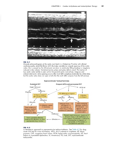
FIG 4.1
M-mode echocardiogram at the aortic root level in a Doberman Pinscher with dilated
cardiomyopathy atrial fibrillation (AF) illustrates variable (or absent) opening of the aortic
valve; this is caused by the AF-induced variation in ventricular filling leading to irregularly
reduced stroke volume. Variable-intensity pulses and pulse deficits result. The motion of
two aortic valve leaflets is seen within the parallel aortic root echoes. Irregular and
abbreviated aortic valve opening occurs after most QRS complexes (indicated by white dots),
but the aortic valve does not open at all after the sixth QRS complex from the left (arrow).
Supraventricular Tachyarrhythmias
Sustained SVT Frequent APCs and paroxysmal SVT
Vagal maneuver HCM cat
±
Ineffective
Effective
PO diltiazem or digoxin
IV fluid to support β-blocker
BP (caution if CHF) or diltiazem
Ineffective Effective
Ineffective
Effective
↑ diltiazem dose, /or Treat underlying
IV diltiazem; repeat add digoxin; or disease; continue
Treat underlying if necessary try β-blocker, or PO diltiazem /or
disease; monitor; other drug, or drug digoxin, or other
consider PO combination (see text) effective drug or
diltiazem or other Effective combination
Ineffective
Repeat vagal maneuver; maximize IV
diltiazem dose; if still ineffective, try
Treat underlying disease;
continue PO diltiazem or other Effective other agents: IV β-blocker, or lidocaine,
appropriate drug; monitor or amiodarone (or digoxin), or
procainamide, or PO sotolol, or
propafenone (see text)
FIG 4.2
A therapeutic approach to supraventricular tachyarrhythmias. See Table 4.2 for drug
doses and text for more information. APCs, Atrial premature complexes; BP, blood
pressure; CHF, congestive heart failure; HCM, hypertrophic cardiomyopathy; HF, heart
failure or myocardial dysfunction; IV, intravenous; PO, oral; SVT, supraventricular
tachycardia.