
Page 233 - Small Animal Internal Medicine, 6th Edition
P. 233
CHAPTER 10 Pulmonary Hypertension and Heartworm Disease 205
Antibody tests. HW antibody (Ab) tests are used to the Ag used in making the test that is too low to be detected.
screen for feline HW exposure. They are relatively sensitive When clinical findings suggest HWD or HARD but the Ab
VetBooks.ir but not specific for adult HWs. The Ab tests detect circulat- test is negative, serologic testing should be repeated using a
different Ab test and an HW Ag test. Thoracic radiographs
ing Abs against a surface protein of male or female L 4 larvae.
These Ab tests have minimal to no cross-reactivity with GI
parasitic infections. Ab tests provide greater sensitivity than and an echocardiogram also are recommended. The Ab test
could be repeated in a few months, as well.
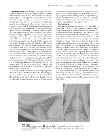
Ag tests because larvae of either sex can provoke a host Radiography
immune response, and Ab tests become positive during the Radiographic findings that suggest HWD are similar to
time frame when cats typically experience clinical signs of those seen in dogs, including pulmonary artery enlarge-
HARD. Serum Ab is detected as early as 60 days after infec- ment with or without visible tortuosity and pruning, RV
tion, although different Ab tests vary in sensitivity to dif- or generalized cardiac enlargement, and diffuse or focal
ferent larval stages. A positive Ab test indicates exposure to pulmonary bronchointerstitial infiltrates (Fig. 10.4). Pul-
migrating larvae and adults, not necessarily the presence of monary hyperinflation sometimes is evident, similar to cats
adult HWs. It is estimated that approximately 50% of Ab- with asthma. The pulmonary artery and right heart changes
positive but Ag-negative cats develop HARD, whereas only typically are more subtle in cats than in dogs. Radio-
10% to 20% develop a mature HW infection. When an Ab graphic abnormalities occur in approximately 50% of cats
test is positive, other evidence should be sought to support with HWD, and may not correlate with severity of clinical
a diagnosis of mature HW infection (and thus true HWD). signs or results of serologic tests. Pulmonary artery disten-
This can include a positive HW Ag test or findings consistent tion may be greatest within the first 7 months of infection.
with HWD on thoracic radiography or echocardiography. Caudal lobar arteries are most frequently abnormal, and are
The concentration of Ab does not appear to correlate well seen best on the DV view. The right caudal lobar artery
with an individual cat’s worm burden nor with the severity can be more prominent; however, a left caudal pulmonary
of clinical disease or radiographic signs, although high Ab artery greater than or equal to 1.6 times the width of the
titers are associated with HW death. Antibodies persist for ninth rib at the ninth intercostal space reportedly is the
6 to 12 months and decrease as HWs mature, such that a cat most discriminating radiographic finding for separating
with an adult HW that has persisted over 12 months may HW-infected from noninfected cats. The main pulmonary
be Ag-positive and Ab-negative. HW Abs are not protective artery segment is not usually visible on DV or ventrodorsal
against future HW infection. views in cats because its location is more medial than it
False-negative Ab tests also occur fairly frequently (up to is in dogs. Marked right heart enlargement is more likely
10%-15% of cases). Therefore a negative HW Ab test suggests when signs of right-sided CHF (e.g., pleural effusion) exist.
one of the following: (1) the cat has not been exposed to Ascites, which is a rare manifestation of CHF secondary to
HWs, (2) the cat has a HW infection less than 60 days old, the common feline cardiomyopathies, occurs in some cats
or (3) the cat produced a concentration of IgG Ab against with HWD.
A B
FIG 10.4
Lateral (A) and dorsoventral (B) radiographs from a cat with heartworm disease. There
are interstitial infiltrates throughout the lung fields and enlarged pulmonary arteries seen
on both views.