
Page 241 - Small Animal Internal Medicine, 6th Edition
P. 241
CHAPTER 11 Systemic Arterial Hypertension 213
BOX 11.2 likely at systolic BPs greater than 180 mm Hg, although it
can develop at lower pressures.
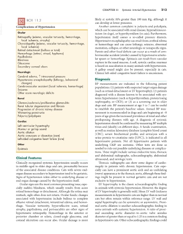
VetBooks.ir Complications of Hypertension which can be associated with renal disease, hyperadrenocor-
Another common complaint is polyuria and polydipsia,
Ocular
Retinopathy (edema, vascular tortuosity, hemorrhage, ticism (in dogs), or hyperthyroidism (in cats). Furthermore,
hypertension itself causes a so-called pressure diuresis.
focal ischemia, atrophy) Hypertensive encephalopathy can result from cerebral edema
Choroidopathy (edema, vascular tortuosity, hemorrhage, or hemorrhage and can cause lethargy, seizures, abnormal
focal ischemia) mentation, collapse, or other neurologic or nonspecific signs.
Retinal detachment (bullous or total) Paresis and other focal defects can occur as a result of cere-
Hemorrhage (retinal, vitreal, hyphema)
Papilledema brovascular accident (stroke) caused by hypertensive arterio-
Blindness lar spasm or hemorrhage. Epistaxis can result from vascular
Glaucoma rupture in the nasal mucosa. A soft, systolic cardiac murmur
Secondary corneal ulcers is heard on auscultation in many animals with hypertension.
A gallop sound might also be present, especially in cats.
Neurologic Clinical left-sided congestive heart failure is uncommon.
Cerebral edema, ↑ intracranial pressure
Hypertensive encephalopathy (lethargy, behavioral Diagnosis
changes) BP measurements are indicated in the following patient
Cerebrovascular accident (focal ischemia, hemorrhage) populations: (1) patients with suspected target organ damage
Seizures (such as retinal detachment or LV hypertrophy); (2) patients
Other acute neurologic deficits
diagnosed with a disease known to be associated with sys-
Renal temic hypertension (such as hyperthyroidism, protein losing
Glomerulosclerosis/proliferative glomerulitis nephropathy, or CKD); or (3) as a screening test in older
Renal tubular degeneration and fibrosis dogs and cats. BP measurement at age 5 to 7 can be useful
Progression of chronic kidney disease to establish the patient’s baseline values. Annual BP mea-
Worsening proteinuria surement is recommended in dogs and cats beginning at ~8
Polyuria/polydipsia years of age given the increased prevalence of renal and other
predisposing diseases with age. A diagnosis of systemic
Cardiac hypertension should be confirmed by measuring BP multiple
Left ventricular hypertrophy times and (ideally) on different days. A fundic examination,
Murmur or gallop sound as well as routine laboratory database (complete blood count
Aortic dilation
Aortic aneurysm or dissection (rare) [CBC]; serum biochemical profile; and urinalysis with a
Left-sided congestive heart failure (rare) urine protein-to-creatinine ratio [UPC]), is indicated in all
hypertensive patients. Not all hypertensive patients with
Other underlying CKD are azotemic. Other tests are done as
Epistaxis needed to rule out possible underlying diseases or complica-
tions. These might include various endocrine tests, thoracic
and abdominal radiographs, echocardiography, abdominal
Clinical Features ultrasound, and serologic tests.
Clinically recognized systemic hypertension usually occurs Thoracic radiographs can show some degree of cardio-
in middle-aged to older dogs and cats, presumably because megaly in patients with chronic hypertension. Cats espe-
of the associated disease conditions. Cats with severe end- cially can have a prominent aortic arch and an undulating
organ disease secondary to hypertension tend to be geriatric. (wavy) appearance to the thoracic aorta, although these find-
Signs of hypertension relate either to underlying disease or ings might be present in normal geriatric cats and are not
end-organ damage caused by the hypertension itself. exclusive to hypertension.
Ocular signs are the most common presenting issue, espe- LV hypertrophy is the classic echocardiographic finding
cially sudden blindness, which usually results from acute in animals with systemic hypertension. However, the degree
retinal hemorrhage or detachment. Although the retina may of LV hypertrophy is generally mild. Mean LV wall thickness
reattach, sight often does not return. Ocular fundic changes measurements in hypertensive cats are higher than in normal
associated with hypertension include bullous to complete cats but often remain within reference range. LV wall and
effusive retinal detachment, intraretinal edema, and hemor- septal hypertrophy can be symmetric or asymmetric. Proxi-
rhage. Vascular tortuosity, hyperreflective scars, retinal mal aortic dilation is another echocardiographic finding in
atrophy, papilledema, and perivasculitis are other signs of some animals with systemic hypertension. A ratio of proxi-
hypertensive retinopathy. Hemorrhage in the anterior or mal ascending aortic diameter–to–aortic valve annulus
posterior chamber or sclera, closed-angle glaucoma, and diameter of greater than or equal to 1.25 is a common finding
corneal ulceration can occur also. Ocular damage is more in hypertensive cats. Other echocardiographic findings could