
Page 358 - Small Animal Internal Medicine, 6th Edition
P. 358
330 PART II Respiratory System Disorders
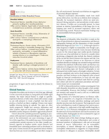
BOX 21.6 the cat’s environment. Seasonal exacerbations are suggestive
of potential allergen exposure.
VetBooks.ir Classification of Feline Bronchial Disease airway obstruction. Cats that are in distress show tachypnea.
Physical examination abnormalities result from small
Bronchial Asthma
Predominant feature: reversible airway obstruction Typically the increased respiratory efforts are more pro-
nounced during expiration, and auscultation reveals expira-
primarily resulting from bronchoconstriction tory wheezes. Crackles are occasionally present. In some
Other common features: hypertrophy of smooth muscle, patients in distress, hyperinflation of the lungs due to air
increased mucus production, eosinophilic inflammation trapping may result in increased inspiratory efforts and
Acute Bronchitis decreased lung sounds. Physical examination findings may
Predominant feature: reversible airway inflammation of be unremarkable between episodes.
short duration (<1-3 months) Diagnosis
Other common features: increased mucus production,
neutrophilic or macrophagic inflammation The diagnosis of idiopathic feline bronchitis is made on the
basis of typical historical, physical examination, and thoracic
Chronic Bronchitis radiographic findings and the elimination of other possible
Predominant feature: chronic airway inflammation (>2-3 differential diagnoses (see Table 21.2). A thorough search for
months) resulting in irreversible damage (e.g., fibrosis) other diagnoses is highly recommended, even though a spe-
Other common features: increased mucus production; cific diagnosis is not commonly found, because identifying
neutrophilic, eosinophilic, or mixed inflammation; a cause for the clinical signs may enable specific treatment
isolation of bacteria or Mycoplasma organisms and even cure of an individual cat. Factors to consider when
causing infection or as nonpathogenic inhabitants;
concurrent bronchial asthma developing a diagnostic plan include the clinical condition
of the cat and the client’s tolerance for expense and risk. Cats
Emphysema that are in respiratory distress or are otherwise in critical
Predominant feature: destruction of bronchiolar and condition should not undergo any stressful testing until their
alveolar walls resulting in enlarged peripheral air condition has stabilized. Sufficiently stable cats that have any
spaces indication of a diagnosis other than idiopathic disease on the
Other common features: cavitary lesions (bullae); result of basis of presenting signs and thoracic radiographs or any
or concurrent with chronic bronchitis subsequent test results require a thorough evaluation. Certain
tests are completely safe, such as fecal testing for pulmonary
Adapted from Moise NS et al.: Bronchopulmonary disease. In parasites, and their inclusion in the diagnostic plan is based
Sherding RG, ed.: The cat: diseases and clinical management,
New York, 1989, Churchill Livingstone. largely on financial considerations. In most cats with signs
of bronchitis, collection of tracheal wash fluid for cytology
and culture and tests for pulmonary parasitism and heart-
progression of signs) can be used to classify the disease in worm disease are recommended.
most cats. A CBC is often performed as a routine screening test. Cats
with idiopathic bronchitis are often thought to have periph-
Clinical Features eral eosinophilia. However, this finding is neither specific
Idiopathic bronchitis can develop in cats of any age, although nor sensitive and cannot be used to rule out or definitively
it most commonly develops in young adult and middle-aged diagnose feline bronchitis.
animals. The major clinical feature is cough or episodic The presence of a bronchial pattern on thoracic radio-
respiratory distress or both. Some clients will confuse cough graphs is supportive of a diagnosis of bronchitis (see Fig.
in cats with attempts to vomit a hairball. Cats that never 20.3). Increased reticular interstitial markings and patchy
produce a hairball are likely coughing. Owners may report alveolar opacities may also be present. The lungs may be seen
audible wheezing during an episode. The signs are often to be overinflated as a result of trapping of air, and occasion-
slowly progressive. Weight loss, anorexia, depression, and ally collapse (i.e., atelectasis) of the right middle lung lobe is
other systemic signs are not present. If systemic signs are seen (see Fig. 20.9). However, radiographs are insensitive for
identified, another diagnosis should be aggressively pursued. the detection of bronchial disease and may be normal in cats
Owners should be carefully questioned regarding an asso- with bronchitis. Radiographs are also scrutinized for signs of
ciation with exposure to potential allergens or irritants. Irri- specific diseases (see Table 21.2).
tants in the environment can cause worsening of signs of Tracheal wash or BAL fluid cytologic findings are gener-
bronchitis regardless of the underlying cause. Environmental ally representative of airway inflammation and consist of
considerations include exposure to new litter (usually per- increased numbers of inflammatory cells and an increased
fumed), cigarette or fireplace smoke, carpet cleaners, and amount of mucus. Inflammation can be eosinophilic,
household items containing perfumes such as deodorant or neutrophilic, or mixed. Although not a specific finding,
hair spray. Clients should also be questioned about whether eosinophilic inflammation is suggestive of a hypersensitiv-
there has been any recent remodeling or any other change in ity response to allergens or parasites. Neutrophils should