
Page 512 - Small Animal Internal Medicine, 6th Edition
P. 512
484 PART III Digestive System Disorders
the dog may be presented for constipation and/or hemato-
chezia. Animals with advanced disease often lose weight. In
VetBooks.ir rare cases there will be infarction of mucosa or vessels with
subsequent ischemia. Cats are rarely affected.
Diagnosis
A serologic test is available (Chapter 27) and appears to have
good sensitivity and specificity. If biopsy is performed, surgi-
cal biopsy or biopsy with rigid biopsy forceps are typically
necessary to obtain submucosa, which is where the organ-
isms are usually found. Eosinophils are typically prominent
in affected tissues. Special stains (e.g., Warthin-Starry) are
often required to find the organism.
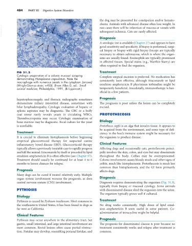
FIG 31.2 Treatment
Cytologic preparation of a colonic mucosal scraping Complete surgical excision is preferred. No medication has
demonstrating Histoplasma capsulatum. Note the
macrophage with numerous yeasts in the cytoplasm (arrows) consistently been effective, although itraconazole or lipid
(Wright-Giemsa stain; ×400). (From Allen D, ed.: Small emulsion amphotericin B plus/minus terbinafine might be
animal medicine, Philadelphia, 1991, JB Lippincott.) temporarily beneficial. Anecdotally, immunotherapy is ben-
eficial in a few patients.
hepatosplenomegaly, and thoracic radiographs sometimes Prognosis
demonstrate miliary interstitial disease, sometimes with The prognosis is poor unless the lesion can be completely
hilar lymphadenopathy. Cytologic evaluation of hepatic or excised.
splenic aspirates may be diagnostic. The CBC or a buffy
coat smear rarely reveals yeasts in circulating WBCs. PROTOTHECOSIS
Thrombocytopenia may occur. Cytologic examination of
bone marrow may be diagnostic. Fecal culture for the yeast Etiology
is unreliable. Prototheca zopfii is an alga that invades tissue. It appears to
be acquired from the environment, and some type of defi-
Treatment ciency in the host’s immune system might be necessary for
It is crucial to eliminate histoplasmosis before beginning the organism to produce disease.
empirical glucocorticoid therapy for suspected canine
inflammatory bowel disease (IBD). Glucocorticoid therapy Clinical Features
typically allows a previously treatable case to rapidly progress Affecting dogs and occasionally cats, protothecosis princi-
and kill the animal. Itraconazole by itself or preceded by lipid pally involves the skin, colon, and eyes but may disseminate
emulsion amphotericin B is often effective (see Chapter 97). throughout the body. Collies may be overrepresented.
Treatment should usually be continued for at least 4 to 6 Colonic involvement causes bloody stools and other signs of
months to lessen chances for relapse. colitis, much like histoplasmosis. Protothecosis is much less
common than histoplasmosis, and the GI form primarily
Prognosis affects dogs.
Many dogs can be cured if treated relatively early. Multiple
organ system involvement worsens the prognosis, as does Diagnosis
central nervous system (CNS) involvement. Diagnosis requires demonstrating the organism (Fig. 31.3),
typically from biopsy or mucosal cytology. Some animals
PYTHIOSIS with disseminated disease shed the organism into the urine.
The organism typically grows well if cultured.
Etiology
Pythiosis is caused by Pythium insidiosum. Most common in Treatment
the southeastern United States, it has been found in dogs as No drug works consistently. High doses of lipid emul-
far west as California. sion amphotericin B seem useful in some patients. Co-
administration of tetracycline might be helpful.
Clinical Features
Pythiosis may occur anywhere in the alimentary tract, but Prognosis
gastric, small intestinal, and large intestinal involvement are The prognosis for disseminated disease is poor because no
most common. Rectal lesions often cause partial obstruc- treatment consistently works and relapse after treatment is
tion. Fistulae may develop, resembling perianal fistulae, and common.