Page 484 - Anatomy and Physiology of Farm Animals, 8th Edition
P. 484

Anatomy of the Female Reproductive System / 469

               appears to take an active part in ovulation,   The uterine tube, like the entire genital
                                                        tract, is invested externally with perito-
               at least to the extent of partially or com-
  VetBooks.ir  pletely enclosing the ovary and directing   neum, which reflects off the organ as a sus-
                                                        pending mesentery. The portion supporting
               the ovum into the uterine tube.
                  The lining of the uterine tube is a much‐  the uterine tube is the mesosalpinx.
               folded mucous membrane covered pri-
               marily with a simple columnar ciliated
               epithelium (Fig.  26‐4B). During estrus   Uterus
               (period of sexual receptivity), the unciliated
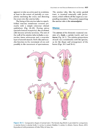
               cells become actively secretory. The rest of   The uterus of the domestic mammal con-
               the wall of the uterine tube includes a con-  sists of a  body, a  cervix (neck), and two
               nective tissue submucosa  and  a  muscular   horns (Fig. 26‐1). The relative proportions
               layer of smooth muscle. Both cilia and mus-  of each vary considerably with the species,
               cles function in the movement of ova and   as do the shape and arrangement of the
               possibly in the movement of spermatozoa.   horns (Figs. 26‐5 and 26‐6).


                                                                1
                                    1


                                                                      2
                                           2
                                     5
                                                               5  4
                                                                    3
                                        4   3


                                          6
                                                                      6


                                         Mare
                                   1                                 Cow

                                                                1
                                          2
                                   5
                                                             5
                                      4  3                        4  2
                                                                    3


                                          6                          6


                                         Sow
                                                                   Bitch
               Figure 26‐5.  Comparative shapes of animal uteri. The female dog (bitch) is provided for comparison.
               1, uterine horn; 2, uterine body; 3, cervix; 4, urinary bladder; 5, ureter; 6, clitoris. Source: Reece, 2015.
               Reproduced with permission of John Wiley & Sons, Inc.
   479   480   481   482   483   484   485   486   487   488   489