
Page 1291 - Veterinary Immunology, 10th Edition
P. 1291
VetBooks.ir
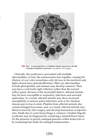
FIG. 39.1 A neutrophil from a Chédiak-Higashi syndrome calf with
enlarged cytoplasmic granules. (Courtesy Dr. H.W. Leipold.)
Clinically, the syndrome is associated with multiple
abnormalities. In hair, the melanosomes fuse together, causing the
dilution of coat color (sometimes only obvious in the newborn) and
light-colored irises (pseudoalbinism). Other eye abnormalities
include photophobia, and animals may develop cataracts. Their
eyes have a red fundic light reflection rather than the normal
yellow-green. Because of the neutrophil defects, affected animals
may be more susceptible to respiratory infections and neonatal
septicemia. As a result, affected animals may show increased
susceptibility to tumors and to infections such as the Aleutian
disease parvovirus in mink. Platelets from affected animals also
contain enlarged lysosomes, and, as a result, affected animals may
bleed excessively after surgery and develop hematomas at injection
sites. Death due to acute hemorrhage is common. Chédiak-Higashi
syndrome may be diagnosed by examining a stained blood smear
for the presence of grossly enlarged granules within leukocytes or
by examining hair shafts for enlarged melanosomes.
1291