
Page 295 - The Veterinary Laboratory and Field Manual 3rd Edition
P. 295
264 Susan C. Cork and Roy Halliwell
Fluorescence In Situ Hybridization [FISH]) and, other viral genera or species. These fixed probes
branched DNA technology (bDNA) that uses are hybridized by target complementary viral
an enzyme-labelled probe plus a chemilumines- genome sequences in the nucleic acid samples
cence-based reaction for signalling). (that are labelled in vitro) extracted from clini-
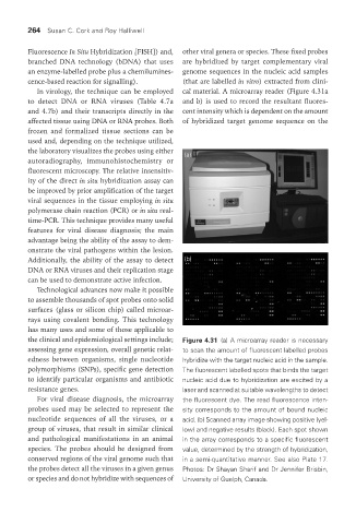
In virology, the technique can be employed cal material. A microarray reader (Figure 4.31a
to detect DNA or RNA viruses (Table 4.7a and b) is used to record the resultant fluores-
and 4.7b) and their transcripts directly in the cent intensity which is dependent on the amount
affected tissue using DNA or RNA probes. Both of hybridized target genome sequence on the
frozen and formalized tissue sections can be
used and, depending on the technique utilized,
the laboratory visualizes the probes using either (a)
autoradiography, immunohistochemistry or
fluorescent microscopy. The relative insensitiv-
ity of the direct in situ hybridization assay can
be improved by prior amplification of the target
viral sequences in the tissue employing in situ
polymerase chain reaction (PCR) or in situ real-
time-PCR. This technique provides many useful
features for viral disease diagnosis; the main
advantage being the ability of the assay to dem-
onstrate the viral pathogens within the lesion.
Additionally, the ability of the assay to detect (b)
DNA or RNA viruses and their replication stage
can be used to demonstrate active infection.
Technological advances now make it possible
to assemble thousands of spot probes onto solid
surfaces (glass or silicon chip) called microar-
rays using covalent bonding. This technology
has many uses and some of those applicable to
the clinical and epidemiological settings include; Figure 4.31 (a) A microarray reader is necessary
assessing gene expression, overall genetic relat- to scan the amount of fluorescent labelled probes
edness between organisms, single nucleotide hybridize with the target nucleic acid in the sample.
polymorphisms (SNPs), specific gene detection The fluorescent labelled spots that binds the target
to identify particular organisms and antibiotic nucleic acid due to hybridization are excited by a
resistance genes. laser and scanned at suitable wavelengths to detect
For viral disease diagnosis, the microarray the fluorescent dye. The read fluorescence inten-
probes used may be selected to represent the sity corresponds to the amount of bound nucleic
nucleotide sequences of all the viruses, or a acid. (b) Scanned array image showing positive (yel-
group of viruses, that result in similar clinical low) and negative results (black). Each spot shown
and pathological manifestations in an animal in the array corresponds to a specific fluorescent
species. The probes should be designed from value, determined by the strength of hybridization,
conserved regions of the viral genome such that in a semi-quantitative manner. See also Plate 17.
the probes detect all the viruses in a given genus Photos: Dr Shayan Sharif and Dr Jennifer Brisbin,
or species and do not hybridize with sequences of University of Guelph, Canada.
Vet Lab.indb 264 26/03/2019 10:25