
Page 302 - The Veterinary Laboratory and Field Manual 3rd Edition
P. 302
Microbiology 271
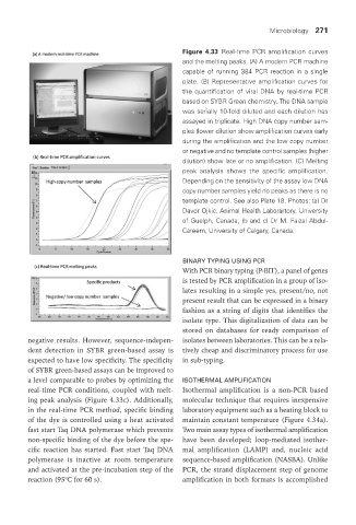
Figure 4.33 Real-time PCR amplification curves
and the melting peaks. (A) A modern PCR machine
capable of running 384 PCR reaction in a single
plate. (B) Representative amplification curves for
the quantification of viral DNA by real-time PCR
based on SYBR Green chemistry. The DNA sample
was serially 10-fold diluted and each dilution has
assayed in triplicate. High DNA copy number sam-
ples (lower dilution show amplification curves early
during the amplification and the low copy number
or negative and no template control samples (higher
dilution) show late or no amplification. (C) Melting
peak analysis shows the specific amplification.
Depending on the sensitivity of the assay low DNA
copy number samples yield no peaks as there is no
template control. See also Plate 18. Photos: (a) Dr
Davor Ojkic, Animal Health Laborartory, University
of Guelph, Canada; (b and c) Dr M. Faizal Abdul-
Careem, University of Calgary, Canada.
bInary tyPInG uSInG Pcr
With PCR binary typing (P-BIT), a panel of genes
is tested by PCR amplification in a group of iso-
lates resulting in a simple yes, present/no, not
present result that can be expressed in a binary
fashion as a string of digits that identifies the
isolate type. This digitalization of data can be
stored on databases for ready comparison of
negative results. However, sequence-indepen- isolates between laboratories. This can be a rela-
dent detection in SYBR green-based assay is tively cheap and discriminatory process for use
expected to have low specificity. The specificity in sub-typing.
of SYBR green-based assays can be improved to
a level comparable to probes by optimizing the ISotHErMaL aMPLIFIcatIon
real-time PCR conditions, coupled with melt- Isothermal amplification is a non-PCR based
ing peak analysis (Figure 4.33c). Additionally, molecular technique that requires inexpensive
in the real-time PCR method, specific binding laboratory equipment such as a heating block to
of the dye is controlled using a heat activated maintain constant temperature (Figure 4.34a).
fast start Taq DNA polymerase which prevents Two main assay types of isothermal amplification
non-specific binding of the dye before the spe- have been developed; loop-mediated isother-
cific reaction has started. Fast start Taq DNA mal amplification (LAMP) and, nucleic acid
polymerase is inactive at room temperature sequence-based amplification (NASBA). Unlike
and activated at the pre-incubation step of the PCR, the strand displacement step of genome
reaction (95°C for 60 s). amplification in both formats is accomplished
Vet Lab.indb 271 26/03/2019 10:25