
Page 169 - Problem-Based Feline Medicine
P. 169
10 – THE CAT WITH TACHYCARDIA, BRADYCARDIA OR AN IRREGULAR RHYTHM 161
Clinical signs related to a fast heart rate (> 280 bpm) If associated with low systemic blood pressure and the
include restlessness, tachypnea, open-mouth breath- cause is not hypovolemia, shock or blood loss, and sys-
ing, poor pulse quality and delayed capillary refill tolic myocardial function is preserved, consider heart
time. rate control with:
● Propranolol at 0.5–1 mg/kg PO q 8–12 hours.
● Atenolol at 6.25–12.5 mg/cat PO q 12 hours.
Diagnosis
● Sotalol at 10–20 mg/cat PO q 12 hours.
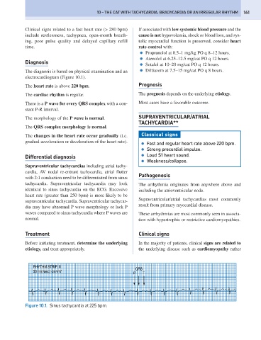
The diagnosis is based on physical examination and an ● Diltiazem at 7.5–15 mg/cat PO q 8 hours.
electrocardiogram (Figure 10.1).
The heart rate is above 220 bpm. Prognosis
The cardiac rhythm is regular. The prognosis depends on the underlying etiology.
There is a P wave for every QRS complex with a con- Most cases have a favorable outcome.
stant P-R interval.
The morphology of the P wave is normal. SUPRAVENTRICULAR/ATRIAL
TACHYCARDIA**
The QRS complex morphology is normal.
The changes in the heart rate occur gradually (i.e. Classical signs
gradual acceleration or deceleration of the heart rate).
● Fast and regular heart rate above 220 bpm.
● Strong precordial impulse.
Differential diagnosis ● Loud S1 heart sound.
● Weakness/collapse.
Supraventricular tachycardias including atrial tachy-
cardia, AV nodal re-entrant tachycardia, atrial flutter
Pathogenesis
with 2:1 conduction need to be differentiated from sinus
tachycardia. Supraventricular tachycardia may look The arrhythmia originates from anywhere above and
identical to sinus tachycardia on the ECG. Excessive including the atrioventricular node.
heart rate (greater than 250 bpm) is more likely to be
Supraventricular/atrial tachycardias most commonly
supraventricular tachycardia. Supraventricular tachycar-
result from primary myocardial disease.
dia may have abnormal P wave morphology or lack P
waves compared to sinus tachycardia where P waves are These arrhythmias are most commonly seen in associa-
normal. tion with hypertrophic or restrictive cardiomyopathies.
Treatment Clinical signs
Before initiating treatment, determine the underlying In the majority of patients, clinical signs are related to
etiology, and treat appropriately. the underlying disease such as cardiomyopathy rather
RHYTHM STRIP: II QRS
50 mm/sec;I cm/mV
P T
Figure 10.1. Sinus tachycardia at 225 bpm.