
Page 1018 - Adams and Stashak's Lameness in Horses, 7th Edition
P. 1018
984 Chapter 9
cow from the herd. After the cut is successfully made, Radiographic abnormalities of the joints may not be
the reins are placed in a relaxed position on the horse’s apparent in young cutting horses with stifle or distal tar
VetBooks.ir during the actual working time. The instinctive ability of short periods of rest combined with NSAIDS and often
sal joint inflammation. Treatment options consist of
neck, and only leg cues are permitted from the rider
IV sodium hyaluronate. Most trainers and owners pre
the working cow horse to contain the individual cow
provides the excitement of competition in cutting. fer to keep the horse in work and elect for intraarticular
During the competition, the horse and rider are judged medication of the medial femorotibial joint and/or both
on how smoothly the cow is separated and kept from the distal intertarsal and tarsometatarsal joints with a
the herd, keeping the cow in the middle of the arena and combination of corticosteroids and sodium hyaluronate.
having the horse work without hand cues from the rider. Many of these horses will also be painful to palpation in
Each contestant gets three cows per performance. The the lumbar region of the back.
rider and horse as a pair begin the competition with a In horses with evidence of soft tissue injuries or radi
score of 72, going up or down from there; a 74–76 is ographic changes, longer periods of rest are usually
a good score. required. Hindlimb proximal suspensory desmitis does
occur in cutting horses. Mild cases with minimal sono
Musculoskeletal Injuries graphic changes respond well to 6–8 weeks of stall rest
combined with local injection of hyaluronic acid and
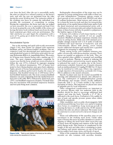
Due to the twisting and quick side‐to‐side movements corticosteroids. Horses with chronic, severe lesions
(Figure 9.36), these horses are plagued with numerous require additional therapies and usually have a guarded
hindlimb, pelvis, and thoracolumbar problems. One ret prognosis for returning to athletic function.
rospective study has documented poor performance and Young cutting horses with hindlimb lameness may
musculoskeletal injuries in 200 horses used solely for cut suffer concurrently with thoracolumbar myositis. These
ting (Dabareiner RM et al., unpublished data). The major horses are painful on palpation of the back. They are
ity of horses were Quarter horses with a median age of 5 often agitated when saddled or mounted and unwilling
years. The most common performance complaint by to stop or perform. Therapy is aimed at reducing the
owners was that the horse would not reverse direction to local inflammation and preventing muscle spasms. Rest
follow the cow (55%), followed by improper stopping is indicated but rarely is an option due to the horse’s
(18%). On physical examination, 70% of horses were performance schedule. Systemic use of muscle relaxants
noted to have thoracolumbar pain upon back palpation. such as methocarbamol (10‐mg/kg PO BID for 10 days)
Eighty percent of horses had peripheral or intra‐articular may be beneficial in mild cases. Others may respond to
diagnostic anesthesia performed to localize the source of a single injection of triamcinolone acetonide (12–16 mg,
lameness. One hundred twenty‐six of 200 horses (63%) IM) in addition to the muscle relaxants.
had hindlimb lameness only. The most common hindlimb Specific localized pain may be treated successfully by
lameness was pain originating from the medial femoroti injecting anti‐inflammatories directly into affected tis
bial joint in the stifle (40%) followed by distal hock pain sues. The combination of methylprednisolone acetate
(20%) and hindlimb suspensory desmitis. Forelimb lame (200–400 mg) and sarapin (50 mL) locally infiltrated
ness was uncommon with proximal suspensory desmitis aseptically with 2‐ to 3.5‐inch needles is the author’s
and foot pain being most common. treatment of choice. The injections are placed into areas
of palpable pain by local infiltration.
Other management considerations are important to
the recovery. Horses with low underrun heels in the
hindfeet should be shod to encourage additional heel
support and raise the heels. Typically, a 2°–3° lift is
indicated in the stock horse. Evaluation of the saddle fit,
type of pad, and specific pressure points when ridden
should be considered.
Other modalities such as pulse electromagnetic field
and ultrasound therapy have been very useful in keeping
these horses in competition. Long warm‐up periods of
ponying the horse at a walk and trot for 30–45 minutes
without the rider are always indicated. The trainer must
be cautioned to avoid overwork and severe fatigue at all
times.
Strain and subluxation of the sacroiliac joint are not
uncommon in the working stock horse. Unlike other
types of horses that incur sacroiliac desmitis as a result of
slipping or falling, the stock horse often incurs this type
of injury from the twisting and rotation of the back and
pelvis during work. This rotation is complicated by the
weight of the tack and rider, who is attempting to main
tain balance and remain stationary on top of the horse
during sudden hard stops, turns, and bursts of speed.
Figure 9.36. Twisting and rotation of the body as the cutting Many of the symptoms observed with thoracolumbar
horse mirrors the movement of the cow. myositis are also common with sacroiliac desmitis.