
Page 1045 - Adams and Stashak's Lameness in Horses, 7th Edition
P. 1045
Occupational‐Related Lameness Conditions 1011
VetBooks.ir
A B
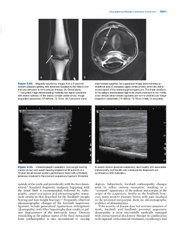
Figure 9.54. Magnetic resonance images from a 9‐year‐old intermediate‐weighted, fat‐suppressed image demonstrating an
western pleasure gelding with lameness localized to the fetlock joint ill‐defined area of increased signal (white arrows) within the mid to
that was refractory to intra‐articular therapy. (A) Dorsal plane, dorsal aspect of the metacarpophalangeal joint. The linear striations
T1‐weighted image demonstrating markedly low signal consistent in the oblique sesamoidean ligaments (most prominent in the medial
with severe sclerosis of the medial condyle (white arrow). Image distal oblique sesamoidean ligament) are normal architecture. Image
acquisition parameters TR 600 ms, TE 12 ms. (B) Transverse plane, acquisition parameters TR 300 ms, TE 40 ms (mildly T2 weighted).
A B
Figure 9.55. Ultrasonographic evaluation (non‐weight bearing to severe chronic proximal suspensory desmopathy with associated
plantar (A‐top) and weight bearing longitudinal (B‐bottom)) of a enthesopathy and fasciitis was subsequently diagnosed and
10‐year‐old all‐around western performance horse with a hindlimb confirmed on MRI evaluation.
lameness localized to the proximal suspensory ligament. Moderate
outside of the circle and periodically with the foot desen degrees. Subjectively, forelimb enthesopathy changes
sitized. Standard diagnostic analgesia beginning with seem to reflect osseous resorption, resulting in a
3
the distal limb is recommended, followed by radio “scooped” appearance of the palmar metacarpus at the
graphic carpal evaluation and ultrasonographic assess origin of the suspensory. Similar to the hindlimb, how
ment similar to that described for the hindlimb (weight ever, many western pleasure horses with pain localized
bearing and non‐weight bearing). Frequently observed to the proximal metacarpus show no ultrasonographic
6,7
ultrasonographic changes of the forelimb suspensory evidence of abnormalities.
ligament include generalized ligamentous enlargement If the severity of disease does not warrant cessation of
(desmopathy) with fiber hypertrophy that results in pal work, forelimb and hindlimb proximal suspensory
mar displacement of the fat/muscle tissue. Osseous desmopathy is most successfully medically managed
remodeling at the palmar aspect of the third metacarpal with extracorporeal shockwave therapy in combination
bone (enthesopathy) is also encountered to varying with regional corticosteroid treatment, cryotherapy, and