
Page 1090 - Adams and Stashak's Lameness in Horses, 7th Edition
P. 1090
1056 Chapter 10
VetBooks.ir
A B
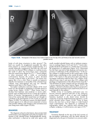
Figure 10.26. Radiographs of the tarsus of two newborn foals on their first day of life. (A) Premature foal. (B) Foal with a normal
gestation period.
laxity of soft tissue structures is often present. The small, rounded cuboidal bones, with or without compres
24
limb can usually be straightened manually but often sion or crushing (Figures 10.26 and 10.27). Serial radio
24
bows inward or outward during weight‐bearing. It is graphs should be performed on a weekly basis to monitor
usually bilaterally symmetrical, and both the carpi and the progression of ossification (Figure 10.2). Ultrasound
tarsi may be affected. When collapse or crushing occurs, has also been reported to help diagnose and monitor pro
this tends to affect the tarsal bones more commonly gression of ossification of the cuboidal bones. In the tar
27
than the carpal bones (Figure 10.27). 12,13 Tarsal collapse sus, collapse is usually located on the cranial aspect of the
is often associated with a sickle or cow‐hocked distal tarsus, contributing to the cranial deviation of the
conformation because the tarsus is often deviated limb. 6,12 The lesions in the tarsus have been described as
cranially (reduced angle to the hock). The tarsus may incomplete ossification with less than 30% collapse of the
1
appear like it has a curb. There is usually no lameness affected bones (type I) and more than 30% collapse with
initially, but lameness is inevitable if the cuboidal bone pinching or fragmentation of the affected bones (type II).
12
collapse went unrecognized and was not clinically Type II lesions are more common and are associated with
apparent until later in life (Figure 10.27). a worse outcome than type I lesions (Figure 10.27). In
12
Incomplete ossification and mild collapse of the tarsal addition, a skeletal ossification index has been proposed to
bones are also thought to predispose to juvenile spavin in classify osseous immaturity in the carpal bones, but it is not
young horses (Figure 10.28). These horses tend to used clinically by the authors. 1
6–8
develop hind lameness and moderate to severe radio Milder forms of incomplete ossification may not
graphic signs of distal tarsal OA at a young age without a become clinically apparent until later in life. However,
history of exercise. Incongruencies or minor malforma young horses (under 2 years of age) with hindlimb
6
tions of the central and third tarsal bones are thought to lameness localized to the distal tarsus should be
contribute to the development of OA at such an early age suspected of having malformation/incongruency of the
(Figure 10.28B). For unknown reasons, the distal intertar tarsal cuboidal bones (Figure 10.28). Some of these
8
8
sal joint appears to be affected most commonly in horses horses also may have a tarsal valgus or sickle‐hocked
with juvenile arthritis, with changes in the tarsometatarsal tarsal conformation. The diagnosis is confirmed by
joint being more subtle (Figure 10.28A). radiographs, which may indicate mild cuboidal bone
collapse, wedging, or malformation, and signs of OA. 7,8
DIAGNOSIS
The diagnosis is based on the history, characteristic limb TREATMENT
conformation, and radiographs indicating incomplete ossi Treatment depends on the age of the foal, severity of
fication of the cuboidal bones. Radiographically, incom the incomplete ossification, and the limbs affected. If
plete ossification is observed as wide joint spaces with diagnosed early, the goal of treatment is to prevent cuboi