
Page 1151 - Adams and Stashak's Lameness in Horses, 7th Edition
P. 1151
Foot Care and Farriery 1117
trimmed foot is fitted with a shoe that extends 1–2 cm heel area to be weight‐bearing but at the same time
beyond the end of the heel and has the break‐over forged decreases the stresses in the DDF muscle tendon unit.
VetBooks.ir the frog and tapering toward the toe to further decrease lameness can be treated by performing a desmotomy of the
Break‐over is applied as described above.
or ground into the shoe starting just dorsal to the apex of
Severe flexural deformities that result in chronic
the stresses on the DDFT.
With a more advanced club foot, the heels should still accessory ligament of the DDFT combined with the appro
be trimmed to load the heels and unload the toe, but priate farriery. 29,46 See “flexural deformities” in Chapter 10
heel elevation must be added to compensate for the for further information regarding this procedure.
shortening of the musculotendinous unit of the DDFT.
This can be determined after the trim by placing the Sheared Heels
trimmed foot palmar to the contralateral limb to observe
whether there is a space between the heels of the foot Sheared heels are a hoof capsule distortion resulting
and the ground. If a space is noted, the author uses a from displacement of one heel bulb proximally relative to
wedge shoe or attaches a wedge pad or a bar wedge to the adjacent heel bulb (Figure 11.36). 18,19,22,30 This disparity
the shoe in order to load the palmar section of the foot between the lateral and medial heel bulb is generally
and to compensate for the shortening of the musculo 0.5 cm or greater. Sheared heels appear to develop as an
tendinous unit (Figure 11.35). This method allows the adaption/distortion of the hoof capsule as a consequence
A B
Figure 11.35. The foot can be placed palmar to the contralateral foot to observe whether there is a space between the heels and the
ground, which corresponds to shortening of the DDFT muscle tendon unit (A). Heel elevation applied to a club foot after trimming (B).
A B
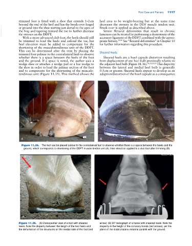
Figure 11.36. (A) Dorsopalmar view of a foot with sheared arrow). (B) DP radiograph of a horse with sheared heels. Note the
heels. Note the disparity between the length of the two heels and disparity in the height of the coronary bands (red arrows), yet the
the deformation of the structures on the medial side of the foot (red plane of the distal phalanx remains parallel with the ground.