
Page 1179 - Adams and Stashak's Lameness in Horses, 7th Edition
P. 1179
Miscellaneous Musculoskeletal Conditions 1145
recovery. First aid measures should be directed toward segments based on anatomy and biomechanical forces
minimizing further damage to the fractured limb and (Figure 12.2). 4,52
VetBooks.ir itate repair. The primary goals of fracture management Phalanges and Distal Metacarpus
maintaining it in a position and condition that will facil
should be as follows :
4
The phalanges and distal metacarpus are probably
1. Prevent damage to neural and vascular elements the most common sites for fractures to occur in
2. Keep the fractured bone from penetrating the skin horses, and biomechanically they are dominated by
and becoming and open fracture or protecting the the angle of the fetlock joint with minimal medial/
4
limb from contamination through any existing skin lateral forces. Therefore, proper splinting techniques
opening should attempt to counteract the bending force at the
3. Stabilize the limb to relieve anxiety that accompanies fetlock. This is achieved by creating dorsal cortical
an uncontrolled limb in a horse alignment (Figure 12.3). A compression bandage
4. Minimize any further damage to the fractured bone combined with a dorsally placed splint or a fiberglass
ends and surrounding soft tissues
cast applied with the limb maintained in a straight
An important purpose of fracture stabilization is to line from the carpus to the hoof provides the optimal
prevent the development of an open fracture. Loss of dorsal cortical alignment (Figure 12.3). Large diame
intact skin coverage over a fracture is thought to pre ter polyvinyl chloride (PVC) pipe cut into thirds along
dispose to infection, especially if internal fixation is its length is a common material used for splints. The
performed. Equine skin is thin and readily penetrated curve of the PVC pipe cradles the bandage and the
by sharp bone fragments, and there is very little soft material is lightweight. Minimal padding (light band
tissue support, such as muscle, below the carpus and age) should be used beneath the splint because frac
tarsus. In general, fractures of the distal (phalanges) ture fragments can move within a large bulky bandage,
and proximal (humerus, ulna, and femur) aspects of and the bulk can sometimes make it difficult for the
the horse’s limbs rarely become open, suggesting that horse to ambulate efficiently. The splint is applied to
fractures involving the metacarpus/metatarsus, radius, the dorsal aspect of the limb from the carpus to the
and tibia are the most critical to proper immobilization hoof and secured with nonelastic tape. A bandage cast
during transport. Immobilization or splinting tech is another alternative—fiberglass casting material
niques depend upon the fracture location and the placed circumferentially over a lightly padded band
52
forces applied to the fracture site. A good rule of age striving to maintain dorsal cortical alignment. To
thumb is that the joint above and below the fracture facilitate application of the splint or bandage cast,
site should be immobilized. In no circumstance should an assistant should hold the forelimb proximal to
the splint end at the same level as the fracture because the carpus and let the limb hang (Figure 12.4).
the splint will act as a fulcrum and further disrupt the Commercially available splints such as the Kimzey
fracture. 52 Leg Saver (Kimzey Welding Works, Woodland, CA;
For immobilization purposes, the forelimb and Figure 12.5) are also available and can be utilized for
hindlimb of the horse can be divided into four functional distal limb fractures.
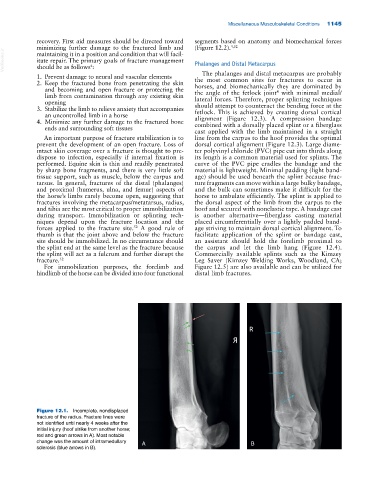
Figure 12.1. Incomplete, nondisplaced
fracture of the radius. Fracture lines were
not identified until nearly 4 weeks after the
initial injury (hoof strike from another horse;
red and green arrows in A). Most notable
change was the amount of intramedullary A B
sclerosis (blue arrows in B).