
Page 1214 - Adams and Stashak's Lameness in Horses, 7th Edition
P. 1214
1180 Chapter 12
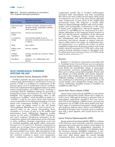
Table 12.5. Alternative explanations for presentations compression usually due to vertebral malformation
with a suspected neurological presentation. associated with osteoarthritis and bony remodeling
VetBooks.ir Alternative, non‐nervous of compression can occur in the same animal, although
that affects the more caudal cervical region. Both causes
static compression is more likely in the middle‐aged
system‐associated explanations
Clinical finding
performance horse. The diagnosis is usually made
based on clinical findings together with cervical radio
Generalized muscle Starvation, emaciation, (intestinal) nutrient graphic and myelographic confirmation of the site(s)
atrophy absorption disorder, paraneoplastic syndrome, of spinal cord compression. Advanced imaging (CT,
chronic myopathies
contrast CT, and MRI) may be used instead of a mye
Regional muscle Lameness‐associated disuse logram depending on the suspected lesion location in
atrophy the neck and the specific capabilities of the advanced
imaging unit. Treatment options include conserva
Low head–neck Nuchal ligament desmitis or rupture, tive management with anti‐inflammatories, exercise
carriage exhaustion, emaciation/starvation, fever, use restriction, and dietary changes or surgical interven
of sedatives tion to either remove the source of compression (dor
sal laminectomy) or to stabilize the area(s) of spinal
Ataxia Fever, sedation
instability/compression. In general, surgery is the most
Stiffness, short Lameness, myopathy widely reported treatment for CVSM and is often indi
strided cated to prevent repetitive trauma to the spinal cord.
Excellent and more extensive reviews of CVSM can be
Hindlimb Laminitis, Chorioptes spp. infestation, fibrotic found in these references. 7,8,12
hypermetria myopathy
Recumbency Exhaustion, colic, rhabdomyolysis, pain, Botulism
laminitis
Botulism is a feed‐borne intoxication associated with
C. botulinum toxin. If several horses are on a property,
botulism usually manifests as a herd outbreak. Botulism
toxin blocks the acetylcholine release at presynaptic
neuromuscular junctions, postganglionic parasympa
SELECT NEUROLOGICAL SYNDROMES thetic nerve endings, and peripheral ganglia. Posture and
AFFECTING THE GAIT gait deficits are noted initially, with muscle fascicula
Cervical Vertebral Stenotic Myelopathy (CVSM) tions and a short‐strided gait, followed by recumbency.
Hallmark findings of botulism are generalized weak
CVSM is probably the most frequent cause of neu ness, resulting in an abnormal gait and recumbency, and
rological gait abnormalities in performance horses. In cranial (motor) nerve dysfunction (tongue tone, masti
general, CVSM is a condition of compression of the cation, swallowing). Excellent reviews of botulism are
cervical spinal cord by malformed cervical vertebrae. available in other texts. 4,14
This focal compression has its greatest impact on white
matter proprioceptive and UMN tracts, resulting in Equine Motor Neuron Disease (EMND)
neurologic deficits. Hallmark findings are generally a
symmetrical ataxia and dysmetria in front limb and Equine motor neuron disease (EMND) is a rare, slowly
hindlimb. The layperson terminology refers to this progressive disease that usually affects only a single ani
condition as “wobbler,” which should be avoided, as a mal in a herd. Although its etiology is unknown, lack of
“wobbler” could be any ataxic horse with numerous antioxidants, particularly vitamin E, has been associ
causes responsible for its clinical presentation. Cervical ated with its development. EMND causes a gradual and
spinal cord compression leading to CVSM is a fre progressive loss of LMN of the entire spinal cord, lead
quent cause of sudden ataxia in the tall breeds ing to signs of symmetrical neurogenic muscle atrophy
(Thoroughbred, Warmblood, etc.) and is typically rec and weakness without ataxia. Horses with EMND are
ognized either in the young, fast‐growing horse described as having a loss of muscle mass in combina
(6 months to 2–3 years of age) or in the middle‐aged tion with a good appetite, and cranial nerves are not
performance horse. Differential diagnoses include affected. 3,11
trauma (to the neck), NAD/EDM, EPM (Americas),
and rarely neoplasia (lymphoma, melanoma). In the Equine Protozoal Myeloencephalitis (EPM)
young horse, spinal cord compression can be a dynamic
or static condition. Dynamic compression occurs due Equine protozoal myeloencephalitis (EPM) is a disease
to vertebral instability and usually affects interverte of the Americas, most commonly caused by a protozoan
bral sites C3–C4 and C4–C5. Dynamic compression is organism, Sarcocystis neurona (and to a lesser degree by
usually seen in younger horses associated with devel Neospora hughesi). The geographical restriction of
opmental orthopedic disease and osteochondrosis at S. neurona is linked to the habitat of its definitive host,
the intervertebral facet joints. Mal‐articulation of the the American opossum (Didelphis virginiana). S. neu-
facet joints can cause abnormal movement between rona oocysts are produced in the opossums’ gastrointes
two vertebrae resulting in spinal cord compression. tinal tract. After horses ingest the oocysts through
Static compression causes continuous spinal cord contaminated feed, sporulation occurs, and asexual