
Page 70 - Adams and Stashak's Lameness in Horses, 7th Edition
P. 70
36 Chapter 1
Long digital extensor m. The tendon of the long digital extensor muscle
extends the length of the metatarsus on the dorsal sur
VetBooks.ir sural n. metatarsus, the tendon of the lateral digital extensor
Caudal cutaneous
face of the cannon bone. At the proximal third of the
muscle joins the long digital extensor tendon. Rarely, the
Lateral
saphenous v. tendon of the lateral digital extensor courses separately
to the proximal phalanx. The angle formed by the con
joined long and lateral digital extensor tendons is occu
pied by the thin, triangular short digital extensor muscle.
Superficial This vestigial muscle originates on the lateral collateral
peroneal n.
ligament of the hock, the lateral tendon of the fibularis
tertius, and the middle extensor retinaculum. It inserts
Lateral digital on the long digital extensor tendon. All digital extensor
extensor m.
muscles are bound down by the distal extensor retinacu
lum in the proximal third of the metatarsus (Figure 1.34).
Proximal Emerging under the distal edge of the distal extensor
extensor retinaculum, the large dorsal metatarsal artery III (“great
retinaculum
metatarsal artery”) runs obliquely to the groove between
the third and fourth metatarsal bones. A very small sat
ellite vein and the lateral dorsal metatarsal nerve run
with the artery. The terminal branch of the caudal cuta
neous sural nerve crosses superficial to the dorsal meta
Middle extensor retinaculum tarsal artery III (Figure 1.34). Distally the artery passes
between the cannon and lateral splint bones near the
Short digital extensor m. button of the splint. Here it continues as the distal per
forating branch, sending branches to the distal deep
Distal extensor retinaculum plantar arch and then dividing into medial and lateral
digital arteries. These continue along the plantar aspect
of the third metatarsal bone in the distal fourth of the
Lateral dorsal metatarsal n. metatarsus.
The lateral dorsal metatarsal nerve remains superfi
cial, courses to the fetlock, and descends on the dorsal
Dorsal metatarsal a. III pastern to terminate in the laminar corium. The medial
dorsal metatarsal nerve supplies sensory fibers to the
hock joint capsule and a motor branch to the short digi
tal extensor muscle. The nerve courses between the long
digital extensor tendon and the second metatarsal bone
Lateral plantar n. to be distributed distally to the pastern and laminar
corium (Figure 1.35).
Superficial plantar arch
Lateral and Medial Aspects
The lateral and medial plantar nerves lie plantar to
Plantar annular their satellite vessels along the dorsal borders of the digi
ligament of tal flexor tendons (Figures 1.34 and 1.35). These nerves
the fetlock supply the lateral, medial, and plantar structures of the
metatarsus. The lateral plantar nerve gives rise to a deep
Proximal digital branch close to the tarsus; this is the parent trunk of the
annular ligament
deeply located lateral and medial plantar metatarsal
Plantar common nerves that pursue courses homologous to the palmar
digital v.III metacarpal nerves in the forelimb. It likewise gives off a
Distal digital sensory branch to the suspensory ligament at this level.
annular ligament At about the mid‐metatarsus, the medial plantar
nerve gives off the communicating branch that angles
laterodistad superficial to the digital flexor tendons to
join the lateral plantar nerve in the distal fourth of the
metatarsus. The communicating branch is generally
smaller than its counterpart in the metacarpus, and it
may be absent.
On each side the very small medial and lateral plantar
arteries arise from the deep plantar arch in the proximal
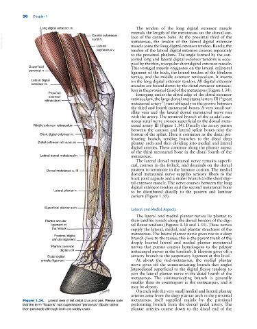
Figure 1.34. Lateral view of left distal crus and pes. Please note metatarsus, itself supplied mainly by the proximal
that the term “fibularis” has superseded “peroneus” (fibular rather perforating branch from the dorsal pedal artery. The
than peroneal) although both are widely used. plantar arteries course down to the distal end of the