
Page 713 - Adams and Stashak's Lameness in Horses, 7th Edition
P. 713
Lameness of the Proximal Limb 679
to identify SCLs as discrete foci of IRU in the tarsus con-
sistent with an active bone remodeling response.
58
VetBooks.ir ize the size and to accurately locate the cystic structure
However, scintigraphy lacks the specificity to character-
and determine communication with the joint. In fact
some cystic lesions are portrayed as a photopenic area,
making them difficult to see these small cystic structures.
Visualization of SCLs in the talus and distal aspect of
the tibia by diagnostic arthroscopy has been described. 69
Depending on the location and extent of the lesion, sur-
gery may be required to prevent cyst enlargement and pro-
mote filling of the lesion with osseous material. Depending
on the cyst location, this can be performed arthroscopi-
cally or via an extra‐articular approach. Additional tech-
niques to improve the healing response in bone and
cartilage may be indicated to preserve articular function.
Malformation of the Distal Tarsal Bones
Malformation of the DT bones can occur in dysma-
ture/immature foals but is most often recognized in pre-
2,3
mature foals. The small cuboidal bones of the carpus
(knee) and tarsus (hock) begin as a cartilage model with
a central area of ossification in utero. As the wave of
ossification progresses, the carpal/tarsal bones change
from a circular central area of ossification into a bony
cube. In normal foals, tarsal bone ossification continues
after birth and is recognized radiographically by a wider
appearance of the joint space due to the presence of
more cartilage compared with an adult horse. If a foal
23
is born prematurely or is dysmature at birth, standing
and normal activity on an incompletely ossified bone
can crush the soft cartilage model. (Figure 5.81A)
3
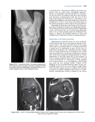
Figure 5.79. Osteoarthritis (OA) of the proximal intertarsal joint Wedging of the dorsal/dorsolateral aspect of the cen-
(PIT) tends to occur in conjunction with and in progression with OA tral/third tarsal bones can occur (Figure 5.81B). These
of the distal tarsal joints. Radiographic changes typically are more foals will often appear to have an angular limb deform-
evident medially as seen on this radiograph (arrows). ity (lateral or medial crushing) in the carpus or a sickle‐
hocked conformation (dorsal crushing) in the tarsus.
A B
Figure 5.80. An SCL in the distal tibia identified on these two MR images (A and B; arrows) communicated with the TC joint in this
horse and caused significant effusion.