
Page 948 - Adams and Stashak's Lameness in Horses, 7th Edition
P. 948
914 Chapter 8
VetBooks.ir
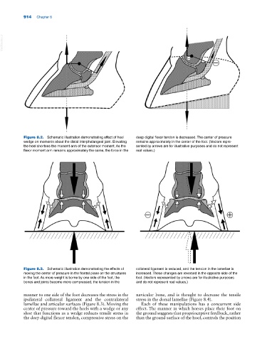
Figure 8.2. Schematic illustration demonstrating effect of heel deep digital flexor tendon is decreased. The center of pressure
wedge on moments about the distal interphalangeal joint. Elevating remains approximately in the center of the foot. (Vectors repre-
the heel shortens the moment arm of the extensor moment. As the sented by arrows are for illustrative purposes and do not represent
flexor moment arm remains approximately the same, the force in the real values.)
Figure 8.3. Schematic illustration demonstrating the effects of collateral ligament is reduced, and the tension in the lamellae is
moving the center of pressure in the frontal plane on the structures increased. These changes are reversed in the opposite side of the
in the foot. As more weight is borne by one side of the foot, the foot. (Vectors represented by arrows are for illustrative purposes
bones and joints become more compressed, the tension in the and do not represent real values.)
manner to one side of the foot decreases the stress in the navicular bone, and is thought to decrease the tensile
ipsilateral collateral ligament and the contralateral stress in the dorsal lamellae (Figure 8.4).
lamellae and articular surfaces (Figure 8.3). Moving the Each of these manipulations has a concurrent side
center of pressure toward the heels with a wedge or any effect. The manner in which horses place their foot on
shoe that functions as a wedge reduces tensile stress in the ground suggests that proprioceptive feedback, rather
the deep digital flexor tendon, compressive stress on the than the ground surface of the hoof, controls the position