
Page 1173 - Equine Clinical Medicine, Surgery and Reproduction, 2nd Edition
P. 1173
1148 CHAPTER 11
VetBooks.ir as atypical. The eyelids, iris, ciliary body, lens, cho- Differential diagnosis
Eyelid lacerations may resemble eyelid colobomas,
roid, retina (including RPE) alone and/or optic disc
may be affected. Colobomas are rare in the horse,
Lens luxation may mimic lens colobomas. Other
but when they do occur they are often seen in com- and synechiae may appear similar to iris colobomas.
bination with other ocular disorders or individually causes of retinal detachment (Table 11.4) must be
as incidental findings in blue-eyed or incompletely ruled out. Glaucoma with optic nerve cupping should
albinotic horses. be easily differentiated from optic nerve colobomas.
Aetiology/pathophysiology Diagnosis
The cause is unknown and the occurrence sporadic. Diagnosis is based on clinical identification of a
Colobomas occur very early in embryogenesis and defect in the eyelid, iris, lens, choroid, retina and/or
are related to defective closure of the embryonic fis- optic nerve that has been present since birth.
sure (typical coloboma) or lack of development of tis-
sue due to a variety of early embryonic stresses or Management
toxins (atypical coloboma). In cases where clinical signs are associated with an
eyelid coloboma, blepharoplasty using advancing
Clinical presentation skin and/or conjunctival flaps can be used to close
Colobomas can vary in appearance from small the defect. Cryoepilation of eyelashes or facial hair
notches in, to an almost complete absence of, tis- contributing to corneal irritation and ocular lubrica-
sue in the eyelid, iris, ciliary body, lens, choroid, tion may also provide relief. There is no therapy for
retina and/or ONH. Trichiasis, leading to keratitis intraocular colobomas; however, in cases of partial
and blepharospasm, may be present secondary to an serous retinal detachment related to fundic colobo-
eyelid coloboma. Dyscoria (abnormal pupil shape) mas, laser therapy may prevent complete detachment.
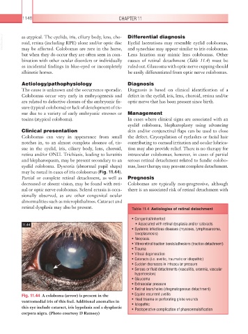
may be noted in cases of iris colobomas (Fig. 11.44).
Partial or complete retinal detachment, as well as Prognosis
decreased or absent vision, may be found with reti- Colobomas are typically non-progressive, although
nal or optic nerve colobomas. Scleral ectasia is occa- there is an associated risk of retinal detachment with
sionally observed, as are other congenital ocular
abnormalities such as microphthalmos. Cataract and
retinal dysplasia may also be present. Table 11.4 Aetiologies of retinal detachment
• Congenital/inherited
11.44 • Associated with retinal dysplasia and/or cataracts
• Systemic infectious diseases (mycoses, lymphosarcoma,
toxoplasmosis)
• Neoplasia
• Vitreoretinal traction bands/adhesions (traction detachment)
• Trauma
• Vitreal degeneration
• Cataracts (i.e. uveitic, traumatic or idiopathic)
• Sudden decreases in intraocular pressure
• Serous or fluid detachments (vasculitis, uraemia, vascular
hypertension)
• Glaucoma
• Extraocular pressure
• Retinal tears/holes (rhegmatogenous detachment)
Fig. 11.44 A coloboma (arrow) is present in the • Equine recurrent uveitis
ventromedial iris of this foal. Additional anomalies in • Head trauma or perforating globe wounds
• Idiopathic
this eye include cataract, iris hypolasia and a dysplastic • Postoperative complication of phaecoemulsification
corpora nigra. (Photo courtesy D Ramsey)